2GRH

M37V mutant of Scapharca dimeric hemoglobin, with CO bound


Experimental Data Snapshot

  • Method: X-RAY DIFFRACTION
  • Resolution: 1.50 Å
  • R-Value Free: 0.194 
  • R-Value Work: 0.186 
  • R-Value Observed: 0.186 

wwPDB Validation   3D Report Full Report


Ligand Structure Quality Assessment 


This is version 1.6 of the entry. See complete history


Literature

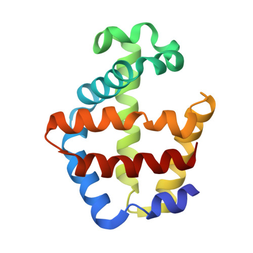
Allosteric action in real time: Time-resolved crystallographic studies of a cooperative dimeric hemoglobin.

Knapp, J.E.Pahl, R.Srajer, V.Royer Jr., W.E.

(2006) Proc Natl Acad Sci U S A 103: 7649-7654

  • DOI: https://doi.org/10.1073/pnas.0509411103
  • Primary Citation of Related Structures:  
    2GRF, 2GRH, 2GRZ

  • PubMed Abstract: 

    Protein allostery provides mechanisms for regulation of biological function at the molecular level. We present here an investigation of global, ligand-induced allosteric transition in a protein by time-resolved x-ray diffraction. The study provides a view of structural changes in single crystals of Scapharca dimeric hemoglobin as they proceed in real time, from 5 ns to 80 micros after ligand photodissociation. A tertiary intermediate structure forms rapidly (<5 ns) as the protein responds to the presence of an unliganded heme within each R-state protein subunit, with key structural changes observed in the heme groups, neighboring residues, and interface water molecules. This intermediate lays a foundation for the concerted tertiary and quaternary structural changes that occur on a microsecond time scale and are associated with the transition to a low-affinity T-state structure. Reversal of these changes shows a considerable lag as a T-like structure persists well after ligand rebinding, suggesting a slow T-to-R transition.


  • Organizational Affiliation

    Department of Biochemistry and Molecular Pharmacology, University of Massachusetts Medical School, Worcester, MA 01655, USA.


Macromolecules
Find similar proteins by:  (by identity cutoff)  |  3D Structure
Entity ID: 1
MoleculeChains Sequence LengthOrganismDetailsImage
Globin-1
A, B
146Anadara inaequivalvisMutation(s): 1 
Gene Names: HBI
UniProt
Find proteins for P02213 (Anadara inaequivalvis)
Explore P02213 
Go to UniProtKB:  P02213
Entity Groups  
Sequence Clusters30% Identity50% Identity70% Identity90% Identity95% Identity100% Identity
UniProt GroupP02213
Sequence Annotations
Expand
  • Reference Sequence
Experimental Data & Validation

Experimental Data

  • Method: X-RAY DIFFRACTION
  • Resolution: 1.50 Å
  • R-Value Free: 0.194 
  • R-Value Work: 0.186 
  • R-Value Observed: 0.186 
  • Space Group: C 1 2 1
Unit Cell:
Length ( Å )Angle ( ˚ )
a = 93.04α = 90
b = 43.93β = 121.95
c = 83.44γ = 90
Software Package:
Software NamePurpose
DENZOdata reduction
SCALEPACKdata scaling
CNSrefinement
CNSphasing

Structure Validation

View Full Validation Report


Ligand Structure Quality Assessment 


Entry History 

Deposition Data

Revision History  (Full details and data files)

  • Version 1.0: 2006-05-09
    Type: Initial release
  • Version 1.1: 2008-05-01
    Changes: Version format compliance
  • Version 1.2: 2011-07-13
    Changes: Version format compliance
  • Version 1.3: 2017-10-18
    Changes: Refinement description
  • Version 1.4: 2018-04-04
    Changes: Data collection
  • Version 1.5: 2021-10-20
    Changes: Database references, Derived calculations
  • Version 1.6: 2023-08-30
    Changes: Data collection, Refinement description