The structure of a d(gcGAACgc) duplex containing two consecutive bulged A residues in both strands suggests a molecular switch
Kondo, J., Sunami, T., Takenaka, A.(2007) Acta Crystallogr D Biol Crystallogr 63: 673-681
- PubMed: 17505105
- DOI: https://doi.org/10.1107/S0907444907012607
- Primary Citation of Related Structures:
2GOT - PubMed Abstract:
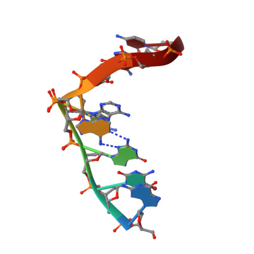
In previous studies, it was reported that DNA fragments with the sequence d(gcGXYAgc) (where X = A or G and Y = A, T or G) form a stable base-intercalated duplex (Bi-duplex) in which the central X and Y residues are not involved in any base-pair interactions but are alternately stacked on each other between the two strands. To investigate the structural stability of the Bi-duplex, the crystal structure of d(gcGAACgc) with a point mutation at the sixth residue of the sequence, d(gcGAAAgc), has been determined. The two strands are associated in an antiparallel fashion to form two types of bulge-containing duplexes (Bc-duplexes), I and II, both of which are quite different from the Bi-duplex of the parent sequence. In both Bc-duplexes, three Watson-Crick G.C base pairs constitute the stem regions at the two ends. The A(4) residues are bulged in to form a pair with the corresponding A(4) residue of the opposite strand in either duplex. The A(4).A(4)* pair formation is correlated to the orientations of the adjacent A(5) residues. A remarkable difference between the two Bc-duplexes is seen at the A(5) residue. In Bc-duplex I, it is flipped out and comes back to interact with the G(3) residue. In Bc-duplex II, the A(5) residue extends outwards to interact with the G(7) residue of the neighbouring Bc-duplex I. These results indicate that trans sugar-edge/Hoogsteen (sheared-type) G(3).A(6)* base pairs are essential in the formation of a Bi-duplex of d(gcGXYAgc). On the other hand, the alternative conformations of the internal loops containing two consecutive bulged A residues suggest molecular switching.
Organizational Affiliation:
Graduate School of Bioscience and Biotechnology, Tokyo Institute of Technology, Yokohama 226-8501, Japan.