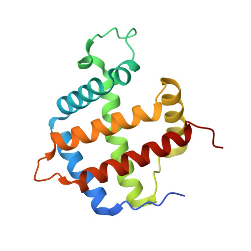
Role of Phenylalanine B10 in Plant Nonsymbiotic Hemoglobins
Smagghe, B.J., Kundu, S., Hoy, J.A., Halder, P., Weiland, T.R., Savage, A., Venugopal, A., Goodman, M., Premer, S., Hargrove, M.S.(2006) Biochemistry 45: 9735-9745
- PubMed: 16893175
- DOI: https://doi.org/10.1021/bi060716s
- Primary Citation of Related Structures:
2GNV, 2GNW - PubMed Abstract:
All plants contain an unusual class of hemoglobins that display bis-histidyl coordination yet are able to bind exogenous ligands such as oxygen. Structurally homologous hexacoordinate hemoglobins (hxHbs) are also found in animals (neuroglobin and cytoglobin) and some cyanobacteria, where they are thought to play a role in free radical scavenging or ligand sensing. The plant hxHbs can be distinguished from the others because they are only weakly hexcacoordinate in the ferrous state, yet no structural mechanism for regulating hexacoordination has been articulated to account for this behavior. Plant hxHbs contain a conserved Phe at position B10 (Phe(B10)), which is near the reversibly coordinated distal His(E7). We have investigated the effects of Phe(B10) mutation on kinetic and equilibrium constants for hexacoordination and exogenous ligand binding in the ferrous and ferric oxidation states. Kinetic and equilibrium constants for hexacoordination and ligand binding along with CO-FTIR spectroscopy, midpoint reduction potentials, and the crystal structures of two key mutant proteins (F40W and F40L) reveal that Phe(B10) is an important regulatory element in hexacoordination. We show that Phe at this position is the only amino acid that facilitates stable oxygen binding to the ferrous Hb and the only one that promotes ligand binding in the ferric oxidation states. This work presents a structural mechanism for regulating reversible intramolecular coordination in plant hxHbs.
Organizational Affiliation:
Department of Biochemistry, Biophysics, and Molecular Biology, Iowa State University, Ames, Iowa 50011, USA.


