2GNU

The crystallization of reaction center from Rhodobacter sphaeroides occurs via a new route


Experimental Data Snapshot

  • Method: X-RAY DIFFRACTION
  • Resolution: 2.20 Å
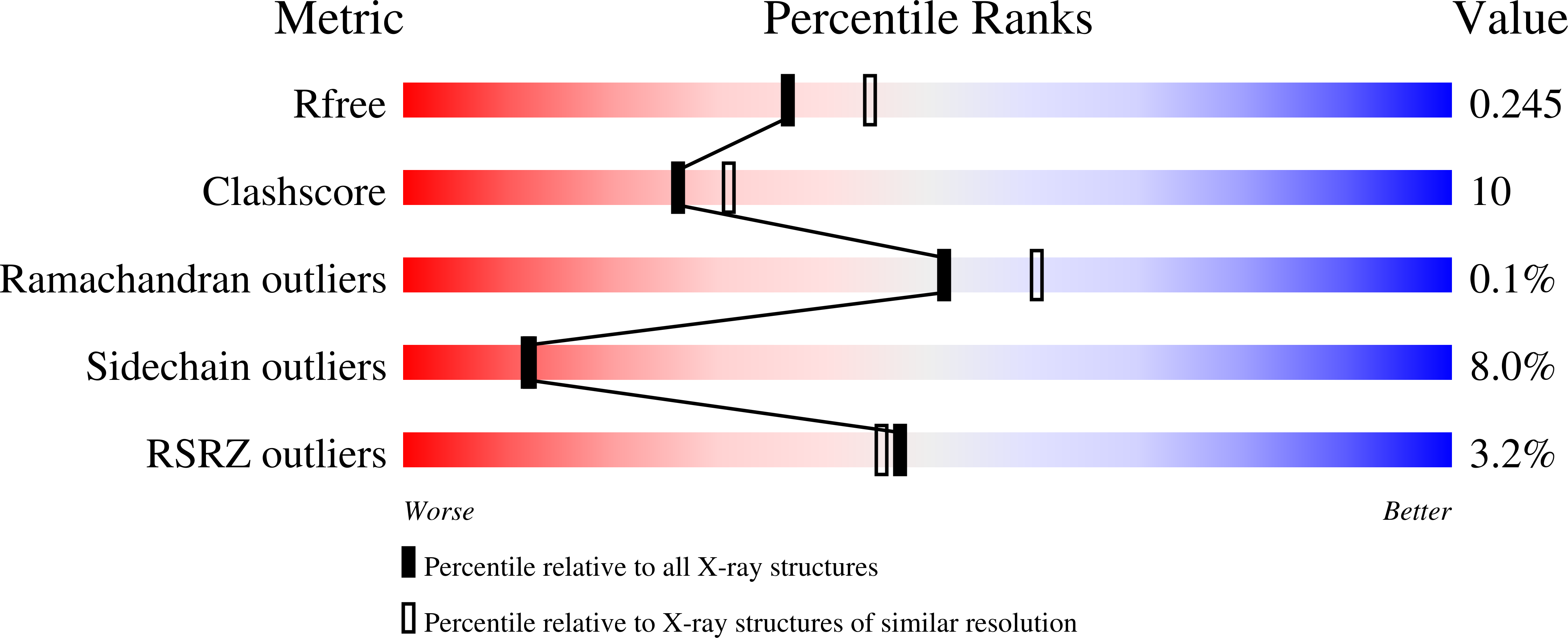
  • R-Value Free: 0.246 
  • R-Value Work: 0.200 

wwPDB Validation   3D Report Full Report


Ligand Structure Quality Assessment 


This is version 1.3 of the entry. See complete history


Literature

Lipidic sponge phase crystallization of membrane proteins

Wadsten, P.Woehri, A.B.Snijder, A.Katona, G.Gardiner, A.T.Cogdell, R.J.Neutze, R.Engstroem, S.

(2006) J Mol Biol 364: 44-53

  • DOI: https://doi.org/10.1016/j.jmb.2006.06.043
  • Primary Citation of Related Structures:  
    2GNU

  • PubMed Abstract: 

    Bicontinuous lipidic cubic phases can be used as a host for growing crystals of membrane proteins. Since the cubic phase is stiff, handling is difficult and time-consuming. Moreover, the conventional cubic phase may interfere with the hydrophilic domains of membrane proteins due to the limited size of the aqueous pores. Here, we introduce a new crystallization method that makes use of a liquid analogue of the cubic phase, the sponge phase. This phase facilitates a considerable increase in the allowed size of aqueous domains of membrane proteins, and is easily generalised to a conventional vapour diffusion crystallisation experiment, including the use of nanoliter drop crystallization robots. The appearance of the sponge phase was confirmed by visual inspection, small-angle X-ray scattering and NMR spectroscopy. Crystals of the reaction centre from Rhodobacter sphaeroides were obtained by a conventional hanging-drop experiment, were harvested directly without the addition of lipase or cryoprotectant, and the structure was refined to 2.2 Angstroms resolution. In contrast to our earlier lipidic cubic phase reaction centre structure, the mobile ubiquinone could be built and refined. The practical advantages of the sponge phase make it a potent tool for crystallization of membrane proteins.


  • Organizational Affiliation

    Department of Chemical and Biological Engineering, Pharmaceutical Technology, Chalmers University of Technology, Göteborg, Sweden.


Macromolecules
Find similar proteins by:  (by identity cutoff)  |  3D Structure
Entity ID: 1
MoleculeChains Sequence LengthOrganismDetailsImage
Reaction center protein H chainA [auth H]235Cereibacter sphaeroidesMutation(s): 0 
Membrane Entity: Yes 
UniProt
Find proteins for Q7B302 (Cereibacter sphaeroides)
Explore Q7B302 
Go to UniProtKB:  Q7B302
Entity Groups  
Sequence Clusters30% Identity50% Identity70% Identity90% Identity95% Identity100% Identity
UniProt GroupQ7B302
Sequence Annotations
Expand
  • Reference Sequence
Find similar proteins by:  (by identity cutoff)  |  3D Structure
Entity ID: 2
MoleculeChains Sequence LengthOrganismDetailsImage
Reaction center protein L chainB [auth L]281Cereibacter sphaeroidesMutation(s): 0 
Membrane Entity: Yes 
UniProt
Find proteins for P0C0Y8 (Cereibacter sphaeroides)
Explore P0C0Y8 
Go to UniProtKB:  P0C0Y8
Entity Groups  
Sequence Clusters30% Identity50% Identity70% Identity90% Identity95% Identity100% Identity
UniProt GroupP0C0Y8
Sequence Annotations
Expand
  • Reference Sequence
Find similar proteins by:  (by identity cutoff)  |  3D Structure
Entity ID: 3
MoleculeChains Sequence LengthOrganismDetailsImage
Reaction center protein M chainC [auth M]300Cereibacter sphaeroidesMutation(s): 0 
Membrane Entity: Yes 
UniProt
Find proteins for P0C0Y9 (Cereibacter sphaeroides)
Explore P0C0Y9 
Go to UniProtKB:  P0C0Y9
Entity Groups  
Sequence Clusters30% Identity50% Identity70% Identity90% Identity95% Identity100% Identity
UniProt GroupP0C0Y9
Sequence Annotations
Expand
  • Reference Sequence
Small Molecules
Ligands 7 Unique
IDChains Name / Formula / InChI Key2D Diagram3D Interactions
CDL
Query on CDL

Download Ideal Coordinates CCD File 
O [auth M]CARDIOLIPIN
C81 H156 O17 P2
XVTUQDWPJJBEHJ-KZCWQMDCSA-L
BCL
Query on BCL

Download Ideal Coordinates CCD File 
E [auth L],
F [auth L],
K [auth M],
L [auth M]
BACTERIOCHLOROPHYLL A
C55 H74 Mg N4 O6
DSJXIQQMORJERS-AGGZHOMASA-M
BPH
Query on BPH

Download Ideal Coordinates CCD File 
J [auth M],
M
BACTERIOPHEOPHYTIN A
C55 H76 N4 O6
KWOZSBGNAHVCKG-SZQBJALDSA-N
U10
Query on U10

Download Ideal Coordinates CCD File 
G [auth L],
N [auth M]
UBIQUINONE-10
C59 H90 O4
ACTIUHUUMQJHFO-UPTCCGCDSA-N
LDA
Query on LDA

Download Ideal Coordinates CCD File 
D [auth H]LAURYL DIMETHYLAMINE-N-OXIDE
C14 H31 N O
SYELZBGXAIXKHU-UHFFFAOYSA-N
FE2
Query on FE2

Download Ideal Coordinates CCD File 
H [auth M]FE (II) ION
Fe
CWYNVVGOOAEACU-UHFFFAOYSA-N
CL
Query on CL

Download Ideal Coordinates CCD File 
I [auth M]CHLORIDE ION
Cl
VEXZGXHMUGYJMC-UHFFFAOYSA-M
Experimental Data & Validation

Experimental Data

  • Method: X-RAY DIFFRACTION
  • Resolution: 2.20 Å
  • R-Value Free: 0.246 
  • R-Value Work: 0.200 
  • Space Group: P 42 21 2
Unit Cell:
Length ( Å )Angle ( ˚ )
a = 100.422α = 90
b = 100.422β = 90
c = 235.412γ = 90
Software Package:
Software NamePurpose
SCALAdata scaling
MOLREPphasing
REFMACrefinement
PDB_EXTRACTdata extraction
MOSFLMdata reduction
CCP4data scaling

Structure Validation

View Full Validation Report


Ligand Structure Quality Assessment 


Entry History 

Deposition Data

Revision History  (Full details and data files)

  • Version 1.0: 2006-11-07
    Type: Initial release
  • Version 1.1: 2008-05-01
    Changes: Version format compliance
  • Version 1.2: 2011-07-13
    Changes: Non-polymer description, Version format compliance
  • Version 1.3: 2023-10-25
    Changes: Data collection, Database references, Derived calculations, Refinement description