2GNN

Crystal Structure of the Orf Virus NZ2 Variant of VEGF-E


Experimental Data Snapshot

  • Method: X-RAY DIFFRACTION
  • Resolution: 2.30 Å
  • R-Value Free: 0.247 
  • R-Value Work: 0.223 
  • R-Value Observed: 0.224 

wwPDB Validation   3D Report Full Report


Ligand Structure Quality Assessment 


This is version 1.4 of the entry. See complete history


Literature

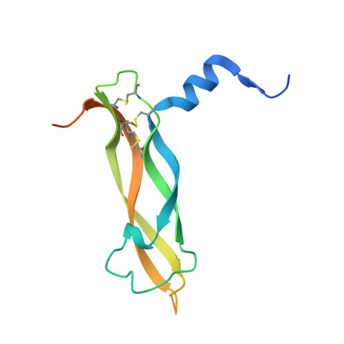
Crystal structure of the Orf virus NZ2 variant of vascular endothelial growth factor-E. Implications for receptor specificity.

Pieren, M.Prota, A.E.Ruch, C.Kostrewa, D.Wagner, A.Biedermann, K.Winkler, F.K.Ballmer-Hofer, K.

(2006) J Biol Chem 281: 19578-19587

  • DOI: https://doi.org/10.1074/jbc.M601842200
  • Primary Citation of Related Structures:  
    2GNN

  • PubMed Abstract: 

    Mammalian vascular endothelial growth factors constitute a family of polypeptides, vascular endothelial growth factor (VEGF)-A, -B, -C, -D and placenta growth factor (PlGF), that regulate blood and lymphatic vessel development. VEGFs bind to three types of receptor tyrosine kinases, VEGF receptors 1, 2, and 3, that are predominantly expressed on endothelial and some hematopoietic cells. Pox viruses of the Orf family encode highly related proteins called VEGF-E that show only 25-35% amino acid identity with VEGF-A but bind with comparable affinity to VEGFR-2. The crystal structure of VEGF-E NZ2 described here reveals high similarity to the known structural homologs VEGF-A, PlGF, and the snake venoms Vammin and VR-1, which are all homodimers and contain the characteristic cysteine knot motif. Distinct conformational differences are observed in loop L1 and particularly in L3, which contains a highly flexible GS-rich motif that differs from all other structural homologs. Based on our structure, we created chimeric proteins by exchanging selected segments in L1 and L3 with the corresponding sequences from PlGF. Single loop mutants did not bind to either receptor, whereas a VEGF-E mutant in which both L1 and L3 were replaced gained affinity for VEGFR-1, illustrating the possibility to engineer receptor-specific chimeric VEGF molecules. In addition, changing arginine 46 to isoleucine in L1 significantly increased the affinity of VEGF-E for both VEGF receptors.


  • Organizational Affiliation

    Molecular Cell Biology, Laboratory of Biomolecular Research, Paul Scherrer Institut, Villigen, Switzerland.


Macromolecules
Find similar proteins by:  (by identity cutoff)  |  3D Structure
Entity ID: 1
MoleculeChains Sequence LengthOrganismDetailsImage
Vascular endothelial growth factor homolog
A, B, C, D
127Orf virus (strain NZ2)Mutation(s): 0 
UniProt
Find proteins for P52584 (Orf virus (strain NZ2))
Explore P52584 
Go to UniProtKB:  P52584
Entity Groups  
Sequence Clusters30% Identity50% Identity70% Identity90% Identity95% Identity100% Identity
UniProt GroupP52584
Sequence Annotations
Expand
  • Reference Sequence
Small Molecules
Ligands 6 Unique
IDChains Name / Formula / InChI Key2D Diagram3D Interactions
NAG
Query on NAG

Download Ideal Coordinates CCD File 
M [auth B],
W [auth D]
2-acetamido-2-deoxy-beta-D-glucopyranose
C8 H15 N O6
OVRNDRQMDRJTHS-FMDGEEDCSA-N
TRS
Query on TRS

Download Ideal Coordinates CCD File 
H [auth A],
U [auth C]
2-AMINO-2-HYDROXYMETHYL-PROPANE-1,3-DIOL
C4 H12 N O3
LENZDBCJOHFCAS-UHFFFAOYSA-O
BEN
Query on BEN

Download Ideal Coordinates CCD File 
G [auth A],
N [auth B],
X [auth D]
BENZAMIDINE
C7 H8 N2
PXXJHWLDUBFPOL-UHFFFAOYSA-N
SO4
Query on SO4

Download Ideal Coordinates CCD File 
F [auth A],
T [auth C]
SULFATE ION
O4 S
QAOWNCQODCNURD-UHFFFAOYSA-L
GOL
Query on GOL

Download Ideal Coordinates CCD File 
I [auth A]
J [auth A]
K [auth A]
L [auth A]
O [auth B]
I [auth A],
J [auth A],
K [auth A],
L [auth A],
O [auth B],
P [auth B],
Q [auth B],
V [auth C]
GLYCEROL
C3 H8 O3
PEDCQBHIVMGVHV-UHFFFAOYSA-N
CL
Query on CL

Download Ideal Coordinates CCD File 
E [auth A],
R [auth C],
S [auth C]
CHLORIDE ION
Cl
VEXZGXHMUGYJMC-UHFFFAOYSA-M
Experimental Data & Validation

Experimental Data

  • Method: X-RAY DIFFRACTION
  • Resolution: 2.30 Å
  • R-Value Free: 0.247 
  • R-Value Work: 0.223 
  • R-Value Observed: 0.224 
  • Space Group: P 41 2 2
Unit Cell:
Length ( Å )Angle ( ˚ )
a = 98.6α = 90
b = 98.6β = 90
c = 240γ = 90
Software Package:
Software NamePurpose
XSCALEdata scaling
REFMACrefinement
PDB_EXTRACTdata extraction
MAR345data collection
SHELXDphasing

Structure Validation

View Full Validation Report


Ligand Structure Quality Assessment 


Entry History 

Deposition Data

Revision History  (Full details and data files)

  • Version 1.0: 2006-05-09
    Type: Initial release
  • Version 1.1: 2008-05-01
    Changes: Version format compliance
  • Version 1.2: 2011-07-13
    Changes: Advisory, Version format compliance
  • Version 1.3: 2017-10-18
    Changes: Refinement description
  • Version 1.4: 2020-07-29
    Type: Remediation
    Reason: Carbohydrate remediation
    Changes: Data collection, Database references, Derived calculations, Structure summary