2GJ4

Structure of rabbit muscle glycogen phosphorylase in complex with ligand


Experimental Data Snapshot

  • Method: X-RAY DIFFRACTION
  • Resolution: 1.60 Å
  • R-Value Free: 0.226 
  • R-Value Work: 0.187 
  • R-Value Observed: 0.189 

wwPDB Validation   3D Report Full Report


Ligand Structure Quality Assessment 


This is version 1.3 of the entry. See complete history


Literature

Novel thienopyrrole glycogen phosphorylase inhibitors: synthesis, in vitro SAR and crystallographic studies.

Whittamore, P.R.Addie, M.S.Bennett, S.N.Birch, A.M.Butters, M.Godfrey, L.Kenny, P.W.Morley, A.D.Murray, P.M.Oikonomakos, N.G.Otterbein, L.R.Pannifer, A.D.Parker, J.S.Readman, K.Siedlecki, P.S.Schofield, P.Stocker, A.Taylor, M.J.Townsend, L.A.Whalley, D.P.Whitehouse, J.

(2006) Bioorg Med Chem Lett 16: 5567-5571

  • DOI: https://doi.org/10.1016/j.bmcl.2006.08.047
  • Primary Citation of Related Structures:  
    2GJ4, 2GM9

  • PubMed Abstract: 

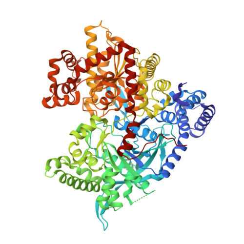
    Two series of novel thienopyrrole inhibitors of recombinant human liver glycogen phosphorylase a (GPa) which are effective in reducing glucose output from rat hepatocytes are described. Representative compounds have been shown to bind at the dimer interface site of the rabbit muscle enzyme by X-ray crystallography.


  • Organizational Affiliation

    AstraZeneca, Alderley Park, Macclesfield, Cheshire, SK10 4TG, UK. paul.whittamore@astrazeneca.com


Macromolecules
Find similar proteins by:  (by identity cutoff)  |  3D Structure
Entity ID: 1
MoleculeChains Sequence LengthOrganismDetailsImage
Glycogen phosphorylase, muscle form824Oryctolagus cuniculusMutation(s): 0 
EC: 2.4.1.1
UniProt
Find proteins for P00489 (Oryctolagus cuniculus)
Explore P00489 
Go to UniProtKB:  P00489
Entity Groups  
Sequence Clusters30% Identity50% Identity70% Identity90% Identity95% Identity100% Identity
UniProt GroupP00489
Sequence Annotations
Expand
  • Reference Sequence
Small Molecules
Ligands 3 Unique
IDChains Name / Formula / InChI Key2D Diagram3D Interactions
2TH
Query on 2TH

Download Ideal Coordinates CCD File 
K [auth A]2-CHLORO-N-[(1R,2R)-1-HYDROXY-2,3-DIHYDRO-1H-INDEN-2-YL]-6H-THIENO[2,3-B]PYRROLE-5-CARBOXAMIDE
C16 H13 Cl N2 O2 S
LRHOLHTVXXSIMG-BXUZGUMPSA-N
PLR
Query on PLR

Download Ideal Coordinates CCD File 
J [auth A](5-HYDROXY-4,6-DIMETHYLPYRIDIN-3-YL)METHYL DIHYDROGEN PHOSPHATE
C8 H12 N O5 P
RBCOYOYDYNXAFA-UHFFFAOYSA-N
SO4
Query on SO4

Download Ideal Coordinates CCD File 
B [auth A]
C [auth A]
D [auth A]
E [auth A]
F [auth A]
B [auth A],
C [auth A],
D [auth A],
E [auth A],
F [auth A],
G [auth A],
H [auth A],
I [auth A]
SULFATE ION
O4 S
QAOWNCQODCNURD-UHFFFAOYSA-L
Binding Affinity Annotations 
IDSourceBinding Affinity
2TH Binding MOAD:  2GJ4 IC50: 319 (nM) from 1 assay(s)
PDBBind:  2GJ4 IC50: 319 (nM) from 1 assay(s)
Experimental Data & Validation

Experimental Data

  • Method: X-RAY DIFFRACTION
  • Resolution: 1.60 Å
  • R-Value Free: 0.226 
  • R-Value Work: 0.187 
  • R-Value Observed: 0.189 
  • Space Group: P 43 21 2
Unit Cell:
Length ( Å )Angle ( ˚ )
a = 128.128α = 90
b = 128.128β = 90
c = 116.346γ = 90
Software Package:
Software NamePurpose
REFMACrefinement
ADSCdata collection
MOSFLMdata reduction
AMoREphasing

Structure Validation

View Full Validation Report


Ligand Structure Quality Assessment 


Entry History 

Deposition Data

Revision History  (Full details and data files)

  • Version 1.0: 2007-02-13
    Type: Initial release
  • Version 1.1: 2008-05-01
    Changes: Version format compliance
  • Version 1.2: 2011-07-13
    Changes: Advisory, Derived calculations, Version format compliance
  • Version 1.3: 2024-04-03
    Changes: Data collection, Database references, Derived calculations, Refinement description