2GE5

EcoRV Restriction Endonuclease C-terminal deletion mutant/GATATC/Ca2+


Experimental Data Snapshot

  • Method: X-RAY DIFFRACTION
  • Resolution: 2.40 Å
  • R-Value Free: 0.284 
  • R-Value Work: 0.223 

wwPDB Validation   3D Report Full Report


This is version 1.3 of the entry. See complete history


Literature

Positively Charged C-Terminal Subdomains of EcoRV Endonuclease: Contributions to DNA Binding, Bending, and Cleavage.

Hiller, D.A.Perona, J.J.

(2006) Biochemistry 45: 11453-11463

  • DOI: https://doi.org/10.1021/bi0606400
  • Primary Citation of Related Structures:  
    2GE5

  • PubMed Abstract: 

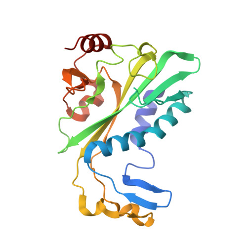
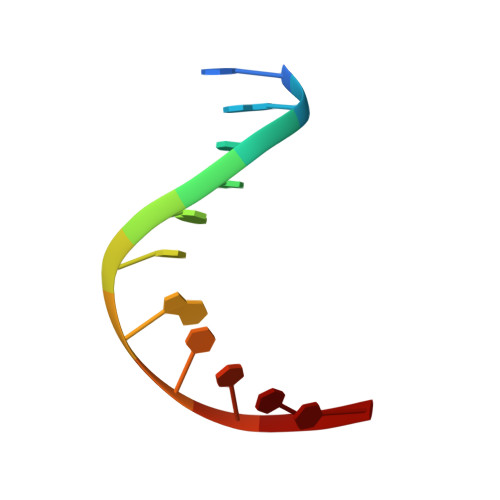
    The carboxy-terminal subdomains of the homodimeric EcoRV restriction endonuclease each bear a net charge of +4 and are positioned on the inner concave surface of the 50 degree DNA bend that is induced by the enzyme. A complete kinetic and structural analysis of a truncated EcoRV mutant lacking these domains was performed to assess the importance of this diffuse charge in facilitating DNA binding, bending, and cleavage. At the level of formation of an enzyme-DNA complex, the association rate for the dimeric mutant enzyme was sharply decreased by 10(3)-fold, while the equilibrium dissociation constant was weakened by nearly 10(6)-fold compared with that of wild-type EcoRV. Thus, the C-terminal subdomains strongly stabilize the enzyme-DNA ground-state complex in which the DNA is known to be bent. Further, the extent of DNA bending as observed by fluorescence resonance energy transfer was also significantly decreased. The crystal structure of the truncated enzyme bound to DNA and calcium ions at 2.4 A resolution reveals that the global fold is preserved and suggests that a divalent metal ion crucial to catalysis is destabilized in the active site. This may explain the 100-fold decrease in the rate of metal-dependent phosphoryl transfer observed for the mutant. These results show that diffuse positive charge associated with the C-terminal subdomains of EcoRV plays a key role in DNA association, bending, and cleavage.


  • Organizational Affiliation

    Department of Chemistry and Biochemistry, University of California, Santa Barbara, California 93106-9510, USA.


Macromolecules

Find similar proteins by:  (by identity cutoff)  |  3D Structure
Entity ID: 2
MoleculeChains Sequence LengthOrganismDetailsImage
Type II restriction enzyme EcoRVC [auth A],
D [auth B]
219Escherichia coliMutation(s): 0 
Gene Names: ecoRVR
EC: 3.1.21.4
UniProt
Find proteins for P04390 (Escherichia coli)
Explore P04390 
Go to UniProtKB:  P04390
Entity Groups  
Sequence Clusters30% Identity50% Identity70% Identity90% Identity95% Identity100% Identity
UniProt GroupP04390
Sequence Annotations
Expand
  • Reference Sequence

Find similar nucleic acids by:  Sequence   |   3D Structure  

Entity ID: 1
MoleculeChains LengthOrganismImage
5'-D(*AP*AP*AP*GP*AP*TP*AP*TP*CP*TP*T)-3'A [auth C],
B [auth D]
11N/A
Sequence Annotations
Expand
  • Reference Sequence
Small Molecules
Ligands 1 Unique
IDChains Name / Formula / InChI Key2D Diagram3D Interactions
CA
Query on CA

Download Ideal Coordinates CCD File 
E [auth A]CALCIUM ION
Ca
BHPQYMZQTOCNFJ-UHFFFAOYSA-N
Experimental Data & Validation

Experimental Data

  • Method: X-RAY DIFFRACTION
  • Resolution: 2.40 Å
  • R-Value Free: 0.284 
  • R-Value Work: 0.223 
  • Space Group: P 43 21 2
Unit Cell:
Length ( Å )Angle ( ˚ )
a = 67.101α = 90
b = 67.101β = 90
c = 259.892γ = 90
Software Package:
Software NamePurpose
SCALAdata scaling
CNSrefinement
PDB_EXTRACTdata extraction
CCP4data scaling
CNSphasing

Structure Validation

View Full Validation Report


Entry History 

Deposition Data

Revision History  (Full details and data files)

  • Version 1.0: 2006-07-04
    Type: Initial release
  • Version 1.1: 2008-05-01
    Changes: Version format compliance
  • Version 1.2: 2011-07-13
    Changes: Version format compliance
  • Version 1.3: 2023-08-30
    Changes: Data collection, Database references, Derived calculations, Refinement description