2GD5

Structural basis for budding by the ESCRTIII factor CHMP3


Experimental Data Snapshot

  • Method: X-RAY DIFFRACTION
  • Resolution: 2.80 Å
  • R-Value Free: 0.301 
  • R-Value Work: 0.259 
  • R-Value Observed: 0.261 

wwPDB Validation   3D Report Full Report


This is version 1.3 of the entry. See complete history


Literature

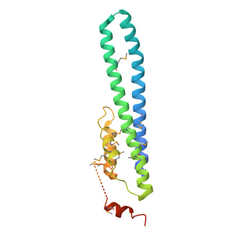
Structural Basis for Budding by the ESCRT-III Factor CHMP3.

Pineda-Molina, E.Ravelli, R.B.Zamborlini, A.Usami, Y.Weissenhorn, W.

(2006) Dev Cell 10: 821-830

  • DOI: https://doi.org/10.1016/j.devcel.2006.03.013
  • Primary Citation of Related Structures:  
    2GD5

  • PubMed Abstract: 

    The vacuolar protein sorting machinery regulates multivesicular body biogenesis and is selectively recruited by enveloped viruses to support budding. Here we report the crystal structure of the human ESCRT-III protein CHMP3 at 2.8 A resolution. The core structure of CHMP3 folds into a flat helical arrangement that assembles into a lattice, mainly via two different dimerization modes, and unilaterally exposes a highly basic surface. The C terminus, the target for Vps4-induced ESCRT disassembly, extends from the opposite side of the membrane targeting region. Mutations within the basic and dimerization regions hinder bilayer interaction in vivo and reverse the dominant-negative effect of a truncated CHMP3 fusion protein on HIV-1 budding. Thus, the final steps in the budding process may include CHMP protein polymerization and lattice formation on membranes by employing different bilayer-recognizing surfaces, a function shared by all CHMP family members.


  • Organizational Affiliation

    European Molecular Biology Laboratory, 6 rue Jules Horowitz, 38042 Grenoble, France.


Macromolecules
Find similar proteins by:  (by identity cutoff)  |  3D Structure
Entity ID: 1
MoleculeChains Sequence LengthOrganismDetailsImage
Charged multivesicular body protein 3
A, B, C, D
179Homo sapiensMutation(s): 12 
Gene Names: CHMP3
UniProt & NIH Common Fund Data Resources
Find proteins for Q9Y3E7 (Homo sapiens)
Explore Q9Y3E7 
Go to UniProtKB:  Q9Y3E7
PHAROS:  Q9Y3E7
GTEx:  ENSG00000115561 
Entity Groups  
Sequence Clusters30% Identity50% Identity70% Identity90% Identity95% Identity100% Identity
UniProt GroupQ9Y3E7
Sequence Annotations
Expand
  • Reference Sequence
Small Molecules
Modified Residues  1 Unique
IDChains TypeFormula2D DiagramParent
MSE
Query on MSE
A, B, C, D
L-PEPTIDE LINKINGC5 H11 N O2 SeMET
Experimental Data & Validation

Experimental Data

  • Method: X-RAY DIFFRACTION
  • Resolution: 2.80 Å
  • R-Value Free: 0.301 
  • R-Value Work: 0.259 
  • R-Value Observed: 0.261 
  • Space Group: P 1
Unit Cell:
Length ( Å )Angle ( ˚ )
a = 33.58α = 68.68
b = 72.57β = 83.61
c = 89.23γ = 76.7
Software Package:
Software NamePurpose
REFMACrefinement
ProDCdata collection
XDSdata scaling
SHELXSphasing

Structure Validation

View Full Validation Report


Entry History 

Deposition Data

Revision History  (Full details and data files)

  • Version 1.0: 2006-06-13
    Type: Initial release
  • Version 1.1: 2008-05-01
    Changes: Version format compliance
  • Version 1.2: 2011-07-13
    Changes: Advisory, Version format compliance
  • Version 1.3: 2017-10-18
    Changes: Refinement description