X-ray crystallographic study of DNA duplex cross-linking: simultaneous binding to two d(CGTACG)2 molecules by a bis(9-aminoacridine-4-carboxamide) derivative.
Hopcroft, N.H., Brogden, A.L., Searcey, M., Cardin, C.J.(2006) Nucleic Acids Res 34: 6663-6672
- PubMed: 17145714
- DOI: https://doi.org/10.1093/nar/gkl930
- Primary Citation of Related Structures:
2GB9 - PubMed Abstract:
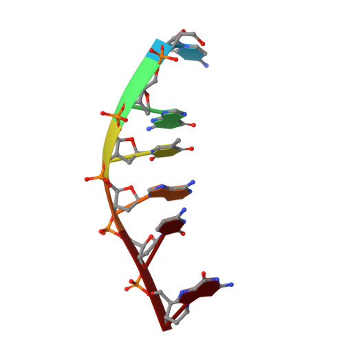
Acridine-4-carboxamides form a class of known DNA mono-intercalating agents that exhibit cytotoxic activity against tumour cell lines due to their ability to inhibit topoisomerases. Previous studies of bis-acridine derivatives have yielded equivocal results regarding the minimum length of linker necessary between the two acridine chromophores to allow bis-intercalation of duplex DNA. We report here the 1.7 A resolution X-ray crystal structure of a six-carbon-linked bis(acridine-4-carboxamide) ligand bound to d(CGTACG)2 molecules by non-covalent duplex cross-linking. The asymmetric unit consists of one DNA duplex containing an intercalated acridine-4-carboxamide chromophore at each of the two CG steps. The other half of each ligand is bound to another DNA molecule in a symmetry-related manner, with the alkyl linker threading through the minor grooves. The two crystallographically independent ligand molecules adopt distinct side chain interactions, forming hydrogen bonds to either O6 or N7 on the major groove face of guanine, in contrast to the semi-disordered state of mono-intercalators bound to the same DNA molecule. The complex described here provides the first structural evidence for the non-covalent cross-linking of DNA by a small molecule ligand and suggests a possible explanation for the inconsistent behaviour of six-carbon linked bis-acridines in previous assays of DNA bis-intercalation.
Organizational Affiliation:
School of Chemistry, University of Reading Whiteknights, Reading, Berkshire RG6 6AD, UK.