


Ligand Epitoping By Proton NMR Chemical Shift Differences
Kline, A.D., Briggs, S.L., Subramaniam, S.To be published.
Experimental Data Snapshot
Entity ID: 1 | |||||
|---|---|---|---|---|---|
| Molecule | Chains | Sequence Length | Organism | Details | Image |
| Cationic trypsin | 223 | Bos taurus | Mutation(s): 0 EC: 3.4.21.4 | 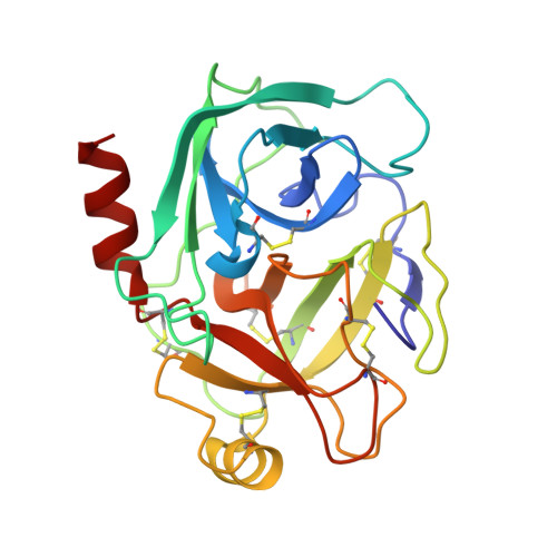 | |
UniProt | |||||
Find proteins for P00760 (Bos taurus) Explore P00760 Go to UniProtKB: P00760 | |||||
Entity Groups | |||||
| Sequence Clusters | 30% Identity50% Identity70% Identity90% Identity95% Identity100% Identity | ||||
| UniProt Group | P00760 | ||||
Sequence AnnotationsExpand | |||||
| |||||
| Ligands 3 Unique | |||||
|---|---|---|---|---|---|
| ID | Chains | Name / Formula / InChI Key | 2D Diagram | 3D Interactions | |
| MI2 Query on MI2 | D [auth A] | 2-(2-METHYLPHENYL)-1H-INDOLE-5-CARBOXIMIDAMIDE C16 H15 N3 YOZKTHGEOJHIDP-UHFFFAOYSA-N |  | ||
| SO4 Query on SO4 | C [auth A] | SULFATE ION O4 S QAOWNCQODCNURD-UHFFFAOYSA-L |  | ||
| CA Query on CA | B [auth A] | CALCIUM ION Ca BHPQYMZQTOCNFJ-UHFFFAOYSA-N |  | ||
| Length ( Å ) | Angle ( ˚ ) |
|---|---|
| a = 54.525 | α = 90 |
| b = 54.525 | β = 90 |
| c = 107.807 | γ = 120 |
| Software Name | Purpose |
|---|---|
| CNX | refinement |
| HKL-2000 | data reduction |
| SCALEPACK | data scaling |
| AMoRE | phasing |
RCSB PDB (citation) is hosted by
RCSB PDB is a member of the

