2G7E

The 1.6 A crystal structure of Vibrio cholerae extracellular endonuclease I


Experimental Data Snapshot

  • Method: X-RAY DIFFRACTION
  • Resolution: 1.60 Å
  • R-Value Free: 0.255 
  • R-Value Work: 0.222 
  • R-Value Observed: 0.224 

wwPDB Validation   3D Report Full Report


This is version 1.3 of the entry. See complete history


Literature

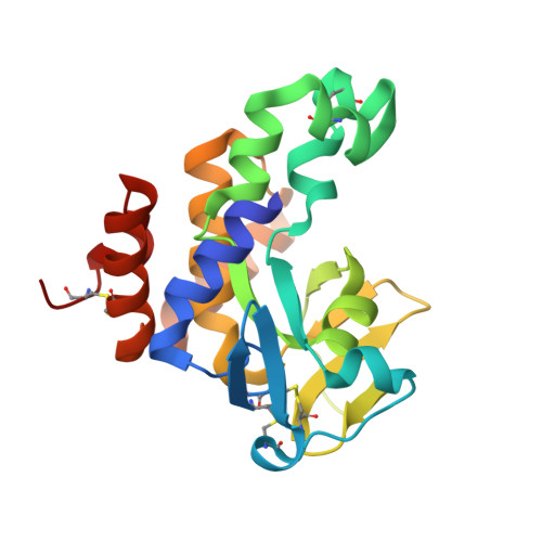
The structure of Vibrio cholerae extracellular endonuclease I reveals the presence of a buried chloride ion.

Altermark, B.Smalas, A.O.Willassen, N.P.Helland, R.

(2006) Acta Crystallogr D Biol Crystallogr 62: 1387-1391

  • DOI: https://doi.org/10.1107/S0907444906034196
  • Primary Citation of Related Structures:  
    2G7E, 2G7F

  • PubMed Abstract: 

    The crystal structure of a periplasmic/extracellular endonuclease from Vibrio cholerae has been solved at low and at neutral pH. Crystals grown at pH 4.6 and 6.9 diffracted to 1.6 A (on BM01A at the ESRF) and 1.95 A (on a rotating-anode generator), respectively. The structures of the endonuclease were compared with the structure of a homologous enzyme in V. vulnificus. The structures of the V. cholerae enzyme at different pH values are essentially identical to each other and to the V. vulnificus enzyme. However, interesting features were observed in the solvent structures. Both V. cholerae structures reveal the presence of a chloride ion completely buried within the core of the protein, with the nearest solvent molecule approximately 7 A away. Magnesium, which is essential for catalysis, is present in the structure at neutral pH, but is absent at low pH, and may partly explain the inactivity of the enzyme at lower pH.


  • Organizational Affiliation

    The Norwegian Structural Biology Centre, Faculty of Science, University of Tromsø, N-9037 Tromsø, Norway.


Macromolecules
Find similar proteins by:  (by identity cutoff)  |  3D Structure
Entity ID: 1
MoleculeChains Sequence LengthOrganismDetailsImage
Endonuclease I211Vibrio choleraeMutation(s): 0 
Gene Names: EndA
EC: 3.1.21.1
UniProt
Find proteins for Q2XSK9 (Vibrio cholerae)
Explore Q2XSK9 
Go to UniProtKB:  Q2XSK9
Entity Groups  
Sequence Clusters30% Identity50% Identity70% Identity90% Identity95% Identity100% Identity
UniProt GroupQ2XSK9
Sequence Annotations
Expand
  • Reference Sequence
Small Molecules
Ligands 1 Unique
IDChains Name / Formula / InChI Key2D Diagram3D Interactions
CL
Query on CL

Download Ideal Coordinates CCD File 
B [auth A]CHLORIDE ION
Cl
VEXZGXHMUGYJMC-UHFFFAOYSA-M
Experimental Data & Validation

Experimental Data

  • Method: X-RAY DIFFRACTION
  • Resolution: 1.60 Å
  • R-Value Free: 0.255 
  • R-Value Work: 0.222 
  • R-Value Observed: 0.224 
  • Space Group: P 21 21 21
Unit Cell:
Length ( Å )Angle ( ˚ )
a = 40.52α = 90
b = 64.16β = 90
c = 75.57γ = 90
Software Package:
Software NamePurpose
SCALAdata scaling
MOLREPphasing
REFMACrefinement
PDB_EXTRACTdata extraction
MOSFLMdata reduction
CCP4data scaling

Structure Validation

View Full Validation Report


Entry History 

Deposition Data

Revision History  (Full details and data files)

  • Version 1.0: 2006-10-31
    Type: Initial release
  • Version 1.1: 2008-05-01
    Changes: Version format compliance
  • Version 1.2: 2011-07-13
    Changes: Version format compliance
  • Version 1.3: 2023-08-30
    Changes: Data collection, Database references, Derived calculations, Refinement description