2G6Y

Crystal structure of the novel green fluorescent protein from marine copepod Pontellina plumata


Experimental Data Snapshot

  • Method: X-RAY DIFFRACTION
  • Resolution: 1.60 Å
  • R-Value Free: 0.218 
  • R-Value Work: 0.162 
  • R-Value Observed: 0.162 

wwPDB Validation   3D Report Full Report


This is version 1.3 of the entry. See complete history


Literature

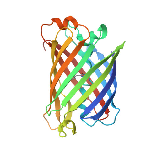
Structural basis for the fast maturation of Arthropoda green fluorescent protein.

Evdokimov, A.G.Pokross, M.E.Egorov, N.S.Zaraisky, A.G.Yampolsky, I.V.Merzlyak, E.M.Shkoporov, A.N.Sander, I.Lukyanov, K.A.Chudakov, D.M.

(2006) EMBO Rep 7: 1006-1012

  • DOI: https://doi.org/10.1038/sj.embor.7400787
  • Primary Citation of Related Structures:  
    2G6X, 2G6Y

  • PubMed Abstract: 

    Since the cloning of Aequorea victoria green fluorescent protein (GFP) in 1992, a family of known GFP-like proteins has been growing rapidly. Today, it includes more than a hundred proteins with different spectral characteristics cloned from Cnidaria species. For some of these proteins, crystal structures have been solved, showing diversity in chromophore modifications and conformational states. However, we are still far from a complete understanding of the origin, functions and evolution of the GFP family. Novel proteins of the family were recently cloned from evolutionarily distant marine Copepoda species, phylum Arthropoda, demonstrating an extremely rapid generation of fluorescent signal. Here, we have generated a non-aggregating mutant of Copepoda fluorescent protein and solved its high-resolution crystal structure. It was found that the protein beta-barrel contains a pore, leading to the chromophore. Using site-directed mutagenesis, we showed that this feature is critical for the fast maturation of the chromophore.


  • Organizational Affiliation

    X-ray crystallography, HCRC, Discovery, Procter&Gamble Pharmaceuticals, 8700 Mason-Montgomery Road, Mason, Ohio 45040, USA.


Macromolecules
Find similar proteins by:  (by identity cutoff)  |  3D Structure
Entity ID: 1
MoleculeChains Sequence LengthOrganismDetailsImage
green fluorescent protein 2
A, B, C, D
217Pontellina plumataMutation(s): 9 
Gene Names: ppluGFP2
UniProt
Find proteins for Q6WV12 (Pontellina plumata)
Explore Q6WV12 
Go to UniProtKB:  Q6WV12
Entity Groups  
Sequence Clusters30% Identity50% Identity70% Identity90% Identity95% Identity100% Identity
UniProt GroupQ6WV12
Sequence Annotations
Expand
  • Reference Sequence
Small Molecules
Modified Residues  1 Unique
IDChains TypeFormula2D DiagramParent
CR2
Query on CR2
A, B, C, D
L-PEPTIDE LINKINGC13 H13 N3 O4GLY, TYR, GLY
Experimental Data & Validation

Experimental Data

  • Method: X-RAY DIFFRACTION
  • Resolution: 1.60 Å
  • R-Value Free: 0.218 
  • R-Value Work: 0.162 
  • R-Value Observed: 0.162 
  • Space Group: P 1 21 1
Unit Cell:
Length ( Å )Angle ( ˚ )
a = 68.007α = 90
b = 97.16β = 96.46
c = 75.153γ = 90
Software Package:
Software NamePurpose
REFMACrefinement
PDB_EXTRACTdata extraction
HKL-2000data reduction
SCALEPACKdata scaling
AMoREphasing

Structure Validation

View Full Validation Report


Entry History 

Deposition Data

Revision History  (Full details and data files)

  • Version 1.0: 2006-03-28
    Type: Initial release
  • Version 1.1: 2008-04-01
    Changes: Version format compliance
  • Version 1.2: 2011-07-13
    Changes: Derived calculations, Version format compliance
  • Version 1.3: 2021-10-20
    Changes: Database references, Derived calculations