2FUF

Crystal structure of the SV40 large T antigen origin-binding domain


Experimental Data Snapshot

  • Method: X-RAY DIFFRACTION
  • Resolution: 1.45 Å
  • R-Value Free: 0.189 
  • R-Value Work: 0.169 
  • R-Value Observed: 0.170 

wwPDB Validation   3D Report Full Report


This is version 1.5 of the entry. See complete history


Literature

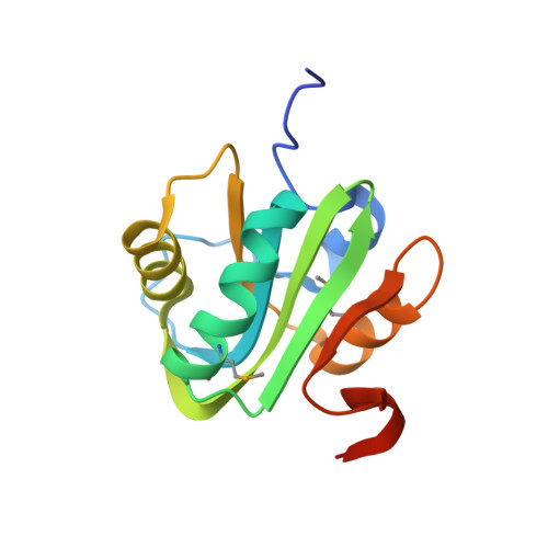
Crystal structure of the simian virus 40 large T-antigen origin-binding domain.

Meinke, G.Bullock, P.A.Bohm, A.

(2006) J Virol 80: 4304-4312

  • DOI: https://doi.org/10.1128/JVI.80.9.4304-4312.2006
  • Primary Citation of Related Structures:  
    2FUF

  • PubMed Abstract: 

    The origins of replication of DNA tumor viruses have a highly conserved feature, namely, multiple binding sites for their respective initiator proteins arranged as inverted repeats. In the 1.45-angstroms crystal structure of the simian virus 40 large T-antigen (T-ag) origin-binding domain (obd) reported herein, T-ag obd monomers form a left-handed spiral with an inner channel of 30 angstroms having six monomers per turn. The inner surface of the spiral is positively charged and includes residues known to bind DNA. Residues implicated in hexamerization of full-length T-ag are located at the interface between adjacent T-ag obd monomers. These data provide a high-resolution model of the hexamer of origin-binding domains observed in electron microscopy studies and allow the obd's to be oriented relative to the hexamer of T-ag helicase domains to which they are connected.


  • Organizational Affiliation

    Tufts University School of Medicine, Department of Biochemistry, 136 Harrison Avenue, Boston, Massachusetts 02111, USA.


Macromolecules
Find similar proteins by:  (by identity cutoff)  |  3D Structure
Entity ID: 1
MoleculeChains Sequence LengthOrganismDetailsImage
Large T antigen134Betapolyomavirus macacaeMutation(s): 2 
Gene Names: SV40 A GENE
UniProt
Find proteins for P03070 (Simian virus 40)
Explore P03070 
Go to UniProtKB:  P03070
Entity Groups  
Sequence Clusters30% Identity50% Identity70% Identity90% Identity95% Identity100% Identity
UniProt GroupP03070
Sequence Annotations
Expand
  • Reference Sequence
Small Molecules
Ligands 1 Unique
IDChains Name / Formula / InChI Key2D Diagram3D Interactions
FLC
Query on FLC

Download Ideal Coordinates CCD File 
B [auth A]CITRATE ANION
C6 H5 O7
KRKNYBCHXYNGOX-UHFFFAOYSA-K
Modified Residues  1 Unique
IDChains TypeFormula2D DiagramParent
MSE
Query on MSE
A
L-PEPTIDE LINKINGC5 H11 N O2 SeMET
Experimental Data & Validation

Experimental Data

  • Method: X-RAY DIFFRACTION
  • Resolution: 1.45 Å
  • R-Value Free: 0.189 
  • R-Value Work: 0.169 
  • R-Value Observed: 0.170 
  • Space Group: P 65
Unit Cell:
Length ( Å )Angle ( ˚ )
a = 83.669α = 90
b = 83.669β = 90
c = 35.88γ = 120
Software Package:
Software NamePurpose
DENZOdata reduction
SCALEPACKdata scaling
PHASERphasing
SOLVEphasing
RESOLVEphasing
REFMACrefinement
PDB_EXTRACTdata extraction

Structure Validation

View Full Validation Report


Entry History 

Deposition Data

Revision History  (Full details and data files)

  • Version 1.0: 2006-08-22
    Type: Initial release
  • Version 1.1: 2008-05-01
    Changes: Version format compliance
  • Version 1.2: 2011-07-13
    Changes: Advisory, Version format compliance
  • Version 1.3: 2017-10-18
    Changes: Refinement description
  • Version 1.4: 2023-08-30
    Changes: Data collection, Database references, Derived calculations, Refinement description
  • Version 1.5: 2023-11-15
    Changes: Data collection