2FI5

Crystal structure of a BPTI variant (Cys38->Ser) in complex with trypsin


Experimental Data Snapshot

  • Method: X-RAY DIFFRACTION
  • Resolution: 1.58 Å
  • R-Value Free: 0.223 
  • R-Value Work: 0.209 
  • R-Value Observed: 0.210 

wwPDB Validation   3D Report Full Report


This is version 1.5 of the entry. See complete history


Literature

Functional and structural roles of the Cys14-Cys38 disulfide of bovine pancreatic trypsin inhibitor.

Zakharova, E.Horvath, M.P.Goldenberg, D.P.

(2008) J Mol Biol 382: 998-1013

  • DOI: https://doi.org/10.1016/j.jmb.2008.07.063
  • Primary Citation of Related Structures:  
    2FI3, 2FI4, 2FI5

  • PubMed Abstract: 

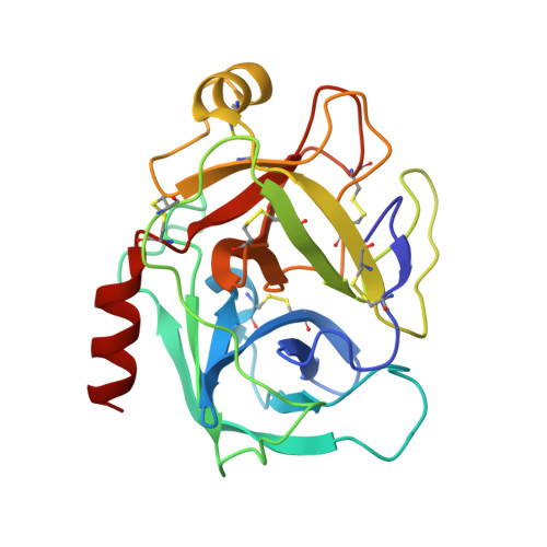
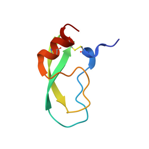
    The disulfide bond between Cys14 and Cys38 of bovine pancreatic trypsin inhibitor lies on the surface of the inhibitor and forms part of the protease-binding region. The functional properties of three variants lacking this disulfide, with one or both of the Cys residues replaced with Ser, were examined, and X-ray crystal structures of the complexes with bovine trypsin were determined and refined to the 1.58-A resolution limit. The crystal structure of the complex formed with the mutant with both Cys residues replaced was nearly identical with that of the complex containing the wild-type protein, with the Ser oxygen atoms positioned to replace the disulfide bond with a hydrogen bond. The two structures of the complexes with single replacements displayed small local perturbations with alternate conformations of the Ser side chains. Despite the absence of the disulfide bond, the crystallographic temperature factors show no evidence of increased flexibility in the complexes with the mutant inhibitors. All three of the variants were cleaved by trypsin more rapidly than the wild-type inhibitor, by as much as 10,000-fold, indicating that the covalent constraint normally imposed by the disulfide contributes to the remarkable resistance to hydrolysis displayed by the wild-type protein. The rates of hydrolysis display an unusual dependence on pH over the range of 3.5-8.0, decreasing at the more alkaline values, as compared with the increased hydrolysis rates for normal substrates under these conditions. These observations can be accounted for by a model for inhibition in which an acyl-enzyme intermediate forms at a significant rate but is rapidly converted back to the enzyme-inhibitor complex by nucleophilic attack by the newly created amino group. The model suggests that a lack of flexibility in the acyl-enzyme intermediate, rather than the enzyme-inhibitor complex, may be a key factor in the ability of bovine pancreatic trypsin inhibitor and similar inhibitors to resist hydrolysis.


  • Organizational Affiliation

    Department of Biology, University of Utah, 257 South 1400 East, Salt Lake City, UT 84112-0840, USA.


Macromolecules
Find similar proteins by:  (by identity cutoff)  |  3D Structure
Entity ID: 1
MoleculeChains Sequence LengthOrganismDetailsImage
Cationic trypsinA [auth E]223Bos taurusMutation(s): 2 
EC: 3.4.21.4
UniProt
Find proteins for P00760 (Bos taurus)
Explore P00760 
Go to UniProtKB:  P00760
Entity Groups  
Sequence Clusters30% Identity50% Identity70% Identity90% Identity95% Identity100% Identity
UniProt GroupP00760
Sequence Annotations
Expand
  • Reference Sequence
Find similar proteins by:  (by identity cutoff)  |  3D Structure
Entity ID: 2
MoleculeChains Sequence LengthOrganismDetailsImage
Pancreatic trypsin inhibitorB [auth I]58Bos taurusMutation(s): 1 
UniProt
Find proteins for P00974 (Bos taurus)
Explore P00974 
Go to UniProtKB:  P00974
Entity Groups  
Sequence Clusters30% Identity50% Identity70% Identity90% Identity95% Identity100% Identity
UniProt GroupP00974
Sequence Annotations
Expand
  • Reference Sequence
Small Molecules
Ligands 4 Unique
IDChains Name / Formula / InChI Key2D Diagram3D Interactions
SO4
Query on SO4

Download Ideal Coordinates CCD File 
E
F [auth E]
O [auth I]
P [auth I]
Q [auth I]
E,
F [auth E],
O [auth I],
P [auth I],
Q [auth I],
R [auth I],
S [auth I],
T [auth I]
SULFATE ION
O4 S
QAOWNCQODCNURD-UHFFFAOYSA-L
EDO
Query on EDO

Download Ideal Coordinates CCD File 
G [auth E]
H [auth E]
I [auth E]
J [auth E]
K [auth E]
G [auth E],
H [auth E],
I [auth E],
J [auth E],
K [auth E],
L [auth E],
M [auth E]
1,2-ETHANEDIOL
C2 H6 O2
LYCAIKOWRPUZTN-UHFFFAOYSA-N
CA
Query on CA

Download Ideal Coordinates CCD File 
D [auth E],
N [auth I]
CALCIUM ION
Ca
BHPQYMZQTOCNFJ-UHFFFAOYSA-N
NA
Query on NA

Download Ideal Coordinates CCD File 
C [auth E]SODIUM ION
Na
FKNQFGJONOIPTF-UHFFFAOYSA-N
Experimental Data & Validation

Experimental Data

  • Method: X-RAY DIFFRACTION
  • Resolution: 1.58 Å
  • R-Value Free: 0.223 
  • R-Value Work: 0.209 
  • R-Value Observed: 0.210 
  • Space Group: I 2 2 2
Unit Cell:
Length ( Å )Angle ( ˚ )
a = 74.726α = 90
b = 81.615β = 90
c = 124.068γ = 90
Software Package:
Software NamePurpose
HKL-2000data collection
SCALEPACKdata scaling
CNSrefinement
HKL-2000data reduction
CNSphasing

Structure Validation

View Full Validation Report


Entry History 

Deposition Data

Revision History  (Full details and data files)

  • Version 1.0: 2006-01-24
    Type: Initial release
  • Version 1.1: 2008-05-01
    Changes: Version format compliance
  • Version 1.2: 2011-07-13
    Changes: Version format compliance
  • Version 1.3: 2011-07-27
    Changes: Database references, Derived calculations, Non-polymer description
  • Version 1.4: 2018-08-29
    Changes: Advisory, Data collection, Source and taxonomy, Structure summary
  • Version 1.5: 2023-08-30
    Changes: Data collection, Database references, Derived calculations, Refinement description