2FGL

An alkali thermostable F/10 xylanase from alkalophilic Bacillus sp. NG-27


Experimental Data Snapshot

  • Method: X-RAY DIFFRACTION
  • Resolution: 2.20 Å
  • R-Value Free: 0.258 
  • R-Value Work: 0.218 
  • R-Value Observed: 0.218 

wwPDB Validation   3D Report Full Report


Ligand Structure Quality Assessment 


This is version 2.1 of the entry. See complete history


Literature

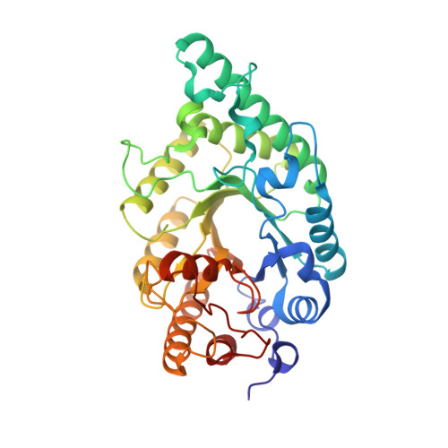
Crystal structures of native and xylosaccharide-bound alkali thermostable xylanase from an alkalophilic Bacillus sp. NG-27: structural insights into alkalophilicity and implications for adaptation to polyextreme conditions.

Manikandan, K.Bhardwaj, A.Gupta, N.Lokanath, N.K.Ghosh, A.Reddy, V.S.Ramakumar, S.

(2006) Protein Sci 15: 1951-1960

  • DOI: https://doi.org/10.1110/ps.062220206
  • Primary Citation of Related Structures:  
    2F8Q, 2FGL

  • PubMed Abstract: 

    Crystal structures are known for several glycosyl hydrolase family 10 (GH10) xylanases. However, none of them is from an alkalophilic organism that can grow in alkaline conditions. We have determined the crystal structures at 2.2 Angstroms of a GH10 extracellular endoxylanase (BSX) from an alkalophilic Bacillus sp. NG-27, for the native and the complex enzyme with xylosaccharides. The industrially important enzyme is optimally active and stable at 343 K and at a pH of 8.4. Comparison of the structure of BSX with those of other thermostable GH10 xylanases optimally active at acidic or close to neutral pH showed that the solvent-exposed acidic amino acids, Asp and Glu, are markedly enhanced in BSX, while solvent-exposed Asn was noticeably depleted. The BSX crystal structure when compared with putative three-dimensional homology models of other extracellular alkalophilic GH10 xylanases from alkalophilic organisms suggests that a protein surface rich in acidic residues may be an important feature common to these alkali thermostable enzymes. A comparison of the surface features of BSX and of halophilic proteins allowed us to predict the activity of BSX at high salt concentrations, which we verified through experiments. This offered us important lessons in the polyextremophilicity of proteins, where understanding the structural features of a protein stable in one set of extreme conditions provided clues about the activity of the protein in other extreme conditions. The work brings to the fore the role of the nature and composition of solvent-exposed residues in the adaptation of enzymes to polyextreme conditions, as in BSX.


  • Organizational Affiliation

    Department of Physics, Indian Institute of Science, Bangalore.


Macromolecules
Find similar proteins by:  (by identity cutoff)  |  3D Structure
Entity ID: 1
MoleculeChains Sequence LengthOrganismDetailsImage
alkaline thermostable endoxylanase
A, B
354Bacillus sp. NG-27Mutation(s): 0 
EC: 3.2.1.8
UniProt
Find proteins for O30700 (Bacillus sp. NG-27)
Explore O30700 
Go to UniProtKB:  O30700
Entity Groups  
Sequence Clusters30% Identity50% Identity70% Identity90% Identity95% Identity100% Identity
UniProt GroupO30700
Sequence Annotations
Expand
  • Reference Sequence
Oligosaccharides

Help

Entity ID: 2
MoleculeChains Length2D Diagram Glycosylation3D Interactions
alpha-D-xylopyranose-(1-1)-alpha-D-xylopyranose
C, D
2N/A
Glycosylation Resources
GlyTouCan:  G77560FK
GlyCosmos:  G77560FK
Small Molecules
Ligands 3 Unique
IDChains Name / Formula / InChI Key2D Diagram3D Interactions
LXC
Query on LXC

Download Ideal Coordinates CCD File 
K [auth B]beta-L-xylopyranose
C5 H10 O5
SRBFZHDQGSBBOR-QTBDOELSSA-N
XYS
Query on XYS

Download Ideal Coordinates CCD File 
E [auth A],
F [auth A],
G [auth A],
J [auth B]
alpha-D-xylopyranose
C5 H10 O5
SRBFZHDQGSBBOR-LECHCGJUSA-N
MG
Query on MG

Download Ideal Coordinates CCD File 
H [auth A],
I [auth A],
L [auth B],
M [auth B]
MAGNESIUM ION
Mg
JLVVSXFLKOJNIY-UHFFFAOYSA-N
Experimental Data & Validation

Experimental Data

  • Method: X-RAY DIFFRACTION
  • Resolution: 2.20 Å
  • R-Value Free: 0.258 
  • R-Value Work: 0.218 
  • R-Value Observed: 0.218 
  • Space Group: P 21 21 21
Unit Cell:
Length ( Å )Angle ( ˚ )
a = 59.188α = 90
b = 84.2β = 90
c = 174.701γ = 90
Software Package:
Software NamePurpose
CNSrefinement
CCP4data scaling
AMoREphasing

Structure Validation

View Full Validation Report


Ligand Structure Quality Assessment 


Entry History 

Deposition Data

Revision History  (Full details and data files)

  • Version 1.0: 2006-09-26
    Type: Initial release
  • Version 1.1: 2008-05-01
    Changes: Version format compliance
  • Version 1.2: 2011-07-13
    Changes: Version format compliance
  • Version 1.3: 2017-10-18
    Changes: Advisory, Refinement description
  • Version 2.0: 2020-07-29
    Type: Remediation
    Reason: Carbohydrate remediation
    Changes: Advisory, Atomic model, Data collection, Derived calculations, Structure summary
  • Version 2.1: 2024-02-14
    Changes: Data collection, Database references, Structure summary