2FE4

The crystal structure of human neuronal Rab6B in its inactive GDP-bound form


Experimental Data Snapshot

  • Method: X-RAY DIFFRACTION
  • Resolution: 2.30 Å
  • R-Value Free: 0.257 
  • R-Value Work: 0.222 
  • R-Value Observed: 0.227 

wwPDB Validation   3D Report Full Report


This is version 1.3 of the entry. See complete history


Literature

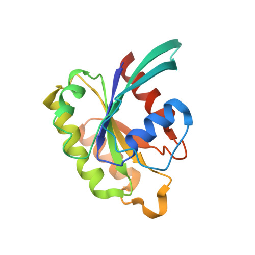
The structure of human neuronal Rab6B in the active and inactive form.

Garcia-Saez, I.Tcherniuk, S.Kozielski, F.

(2006) Acta Crystallogr D Biol Crystallogr 62: 725-733

  • DOI: https://doi.org/10.1107/S0907444906015319
  • Primary Citation of Related Structures:  
    2FE4, 2FFQ

  • PubMed Abstract: 

    The Rab small G-protein family plays important roles in eukaryotes as regulators of vesicle traffic. In Rab proteins, the hydrolysis of GTP to GDP is coupled with association with and dissociation from membranes. Conformational changes related to their different nucleotide states determine their effector specificity. The crystal structure of human neuronal Rab6B was solved in its 'inactive' (with bound MgGDP) and 'active' (MgGTPgammaS-bound) forms to 2.3 and 1.8 A, respectively. Both crystallized in space group P2(1)2(1)2(1), with similar unit-cell parameters, allowing the comparison of both structures without packing artifacts. Conformational changes between the inactive GDP and active GTP-like state are observed mainly in the switch I and switch II regions, confirming their role as a molecular switch. Compared with other Rab proteins, additional changes are observed in the Rab6 subfamily-specific RabSF3 region that might contribute to the specificity of Rab6 for its different effector proteins.


  • Organizational Affiliation

    Laboratoire des Moteurs Moléculaires (LMM), Institut de Biologie Structurale (CEA-CNRS-UJF), 41 Rue Jules Horowitz, 38027 Grenoble CEDEX 01, France.


Macromolecules
Find similar proteins by:  (by identity cutoff)  |  3D Structure
Entity ID: 1
MoleculeChains Sequence LengthOrganismDetailsImage
Ras-related protein Rab-6B171Homo sapiensMutation(s): 0 
Gene Names: RAB6B
EC: 3.6.1
UniProt & NIH Common Fund Data Resources
Find proteins for Q9NRW1 (Homo sapiens)
Explore Q9NRW1 
Go to UniProtKB:  Q9NRW1
PHAROS:  Q9NRW1
GTEx:  ENSG00000154917 
Entity Groups  
Sequence Clusters30% Identity50% Identity70% Identity90% Identity95% Identity100% Identity
UniProt GroupQ9NRW1
Sequence Annotations
Expand
  • Reference Sequence
Experimental Data & Validation

Experimental Data

  • Method: X-RAY DIFFRACTION
  • Resolution: 2.30 Å
  • R-Value Free: 0.257 
  • R-Value Work: 0.222 
  • R-Value Observed: 0.227 
  • Space Group: P 21 21 21
Unit Cell:
Length ( Å )Angle ( ˚ )
a = 36.036α = 90
b = 61.125β = 90
c = 66.634γ = 90
Software Package:
Software NamePurpose
DENZOdata reduction
SCALAdata scaling
AMoREphasing
CNSrefinement
CCP4data scaling

Structure Validation

View Full Validation Report


Entry History 

Deposition Data

Revision History  (Full details and data files)

  • Version 1.0: 2006-07-04
    Type: Initial release
  • Version 1.1: 2008-05-01
    Changes: Version format compliance
  • Version 1.2: 2011-07-13
    Changes: Version format compliance
  • Version 1.3: 2024-02-14
    Changes: Data collection, Database references, Derived calculations