2F93

K Intermediate Structure of Sensory Rhodopsin II/Transducer Complex in Combination with the Ground State Structure


Experimental Data Snapshot

  • Method: X-RAY DIFFRACTION
  • Resolution: 2.00 Å
  • R-Value Free: 0.245 
  • R-Value Work: 0.218 
  • R-Value Observed: 0.218 

wwPDB Validation   3D Report Full Report


This is version 1.4 of the entry. See complete history


Literature

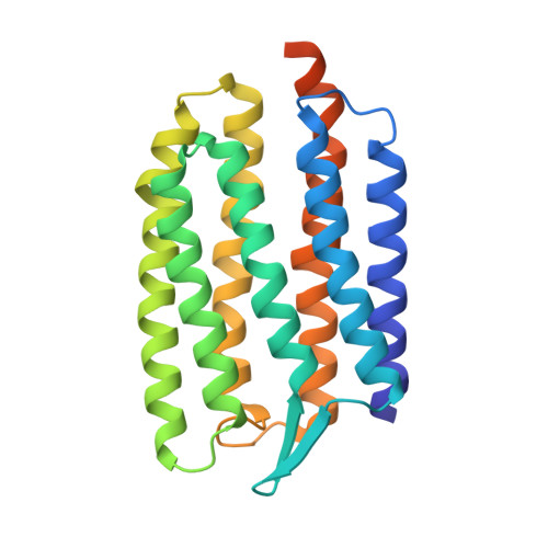
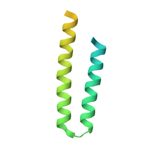
Development of the signal in sensory rhodopsin and its transfer to the cognate transducer.

Moukhametzianov, R.Klare, J.P.Efremov, R.Baeken, C.Goppner, A.Labahn, J.Engelhard, M.Buldt, G.Gordeliy, V.I.

(2006) Nature 440: 115-119

  • DOI: https://doi.org/10.1038/nature04520
  • Primary Citation of Related Structures:  
    2F93, 2F95

  • PubMed Abstract: 

    The microbial phototaxis receptor sensory rhodopsin II (NpSRII, also named phoborhodopsin) mediates the photophobic response of the haloarchaeon Natronomonas pharaonis by modulating the swimming behaviour of the bacterium. After excitation by blue-green light NpSRII triggers, by means of a tightly bound transducer protein (NpHtrII), a signal transduction chain homologous with the two-component system of eubacterial chemotaxis. Two molecules of NpSRII and two molecules of NpHtrII form a 2:2 complex in membranes as shown by electron paramagnetic resonance and X-ray structure analysis. Here we present X-ray structures of the photocycle intermediates K and late M (M2) explaining the evolution of the signal in the receptor after retinal isomerization and the transfer of the signal to the transducer in the complex. The formation of late M has been correlated with the formation of the signalling state. The observed structural rearrangements allow us to propose the following mechanism for the light-induced activation of the signalling complex. On excitation by light, retinal isomerization leads in the K state to a rearrangement of a water cluster that partly disconnects two helices of the receptor. In the transition to late M the changes in the hydrogen bond network proceed further. Thus, in late M state an altered tertiary structure establishes the signalling state of the receptor. The transducer responds to the activation of the receptor by a clockwise rotation of about 15 degrees of helix TM2 and a displacement of this helix by 0.9 A at the cytoplasmic surface.


  • Organizational Affiliation

    Research Centre Jülich, Institute of Structural Biology (IBI-2), 52425 Jülich, Germany.


Macromolecules
Find similar proteins by:  (by identity cutoff)  |  3D Structure
Entity ID: 1
MoleculeChains Sequence LengthOrganismDetailsImage
Sensory rhodopsin II248Natronomonas pharaonisMutation(s): 0 
Gene Names: sop2sopII
Membrane Entity: Yes 
UniProt
Find proteins for P42196 (Natronomonas pharaonis)
Explore P42196 
Go to UniProtKB:  P42196
Entity Groups  
Sequence Clusters30% Identity50% Identity70% Identity90% Identity95% Identity100% Identity
UniProt GroupP42196
Sequence Annotations
Expand
  • Reference Sequence
Find similar proteins by:  (by identity cutoff)  |  3D Structure
Entity ID: 2
MoleculeChains Sequence LengthOrganismDetailsImage
Sensory rhodopsin II transducer122Natronomonas pharaonisMutation(s): 0 
Gene Names: htr2htrII
Membrane Entity: Yes 
UniProt
Find proteins for P42259 (Natronomonas pharaonis)
Explore P42259 
Go to UniProtKB:  P42259
Entity Groups  
Sequence Clusters30% Identity50% Identity70% Identity90% Identity95% Identity100% Identity
UniProt GroupP42259
Sequence Annotations
Expand
  • Reference Sequence
Small Molecules
Ligands 2 Unique
IDChains Name / Formula / InChI Key2D Diagram3D Interactions
BOG
Query on BOG

Download Ideal Coordinates CCD File 
C [auth A]octyl beta-D-glucopyranoside
C14 H28 O6
HEGSGKPQLMEBJL-RKQHYHRCSA-N
RET
Query on RET

Download Ideal Coordinates CCD File 
D [auth A]RETINAL
C20 H28 O
NCYCYZXNIZJOKI-OVSJKPMPSA-N
Experimental Data & Validation

Experimental Data

  • Method: X-RAY DIFFRACTION
  • Resolution: 2.00 Å
  • R-Value Free: 0.245 
  • R-Value Work: 0.218 
  • R-Value Observed: 0.218 
  • Space Group: P 21 21 2
Unit Cell:
Length ( Å )Angle ( ˚ )
a = 124.34α = 90
b = 47.03β = 90
c = 53.82γ = 90
Software Package:
Software NamePurpose
CNSrefinement
MOSFLMdata reduction
CCP4data scaling
CNSphasing

Structure Validation

View Full Validation Report


Entry History 

Deposition Data

Revision History  (Full details and data files)

  • Version 1.0: 2006-03-07
    Type: Initial release
  • Version 1.1: 2008-01-03
    Changes: Version format compliance
  • Version 1.2: 2011-07-13
    Changes: Derived calculations, Version format compliance
  • Version 1.3: 2020-07-29
    Type: Remediation
    Reason: Carbohydrate remediation
    Changes: Data collection, Database references, Derived calculations, Structure summary
  • Version 1.4: 2023-08-30
    Changes: Data collection, Database references, Refinement description, Structure summary