2F2F

Crystal structure of cytolethal distending toxin (CDT) from Actinobacillus actinomycetemcomitans


Experimental Data Snapshot

  • Method: X-RAY DIFFRACTION
  • Resolution: 2.40 Å
  • R-Value Free: 0.251 
  • R-Value Work: 0.221 
  • R-Value Observed: 0.224 

wwPDB Validation   3D Report Full Report


This is version 1.4 of the entry. See complete history


Literature

Variation of loop sequence alters stability of cytolethal distending toxin (CDT): crystal structure of CDT from Actinobacillus actinomycetemcomitans

Yamada, T.Komoto, J.Saiki, K.Konishi, K.Takusagawa, F.

(2006) Protein Sci 15: 362-372

  • DOI: https://doi.org/10.1110/ps.051790506
  • Primary Citation of Related Structures:  
    2F2F

  • PubMed Abstract: 

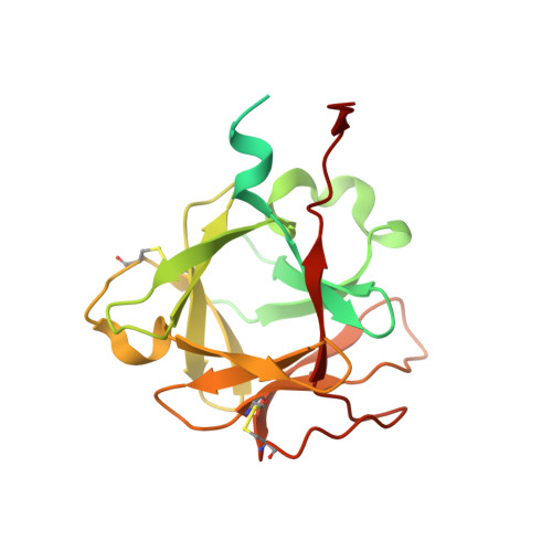
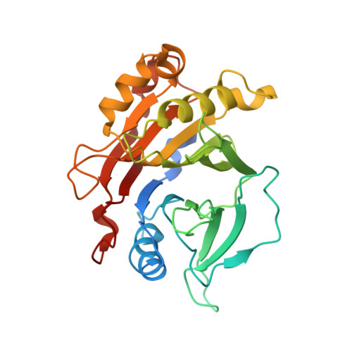
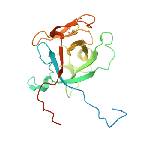
    Cytolethal distending toxin (CDT) secreted by Actinobacillus actinomycetemcomitans induces cell cycle arrest of cultured cells in the G2 phase. The crystal structure of the natural form of A. actinomycetemcomitans DCT (aCDT) has been determined at 2.4 A resolution. aCDT is a heterotrimer consisting of the three subunits, aCdtA, aCdtB, and aCdtC. Two crystallographically independent aCDTs form a dimer through interactions of the aCdtB subunits. The primary structure of aCDT has 94.3% identity with that of Haemophilus ducreyi CDT (hCDT), and the structure of aCDT is quite similar to that of hCDT reconstituted from the three subunits determined recently. However, the molecular packings in the crystal structures of aCDT and hCDT are quite different. A careful analysis of molecular packing suggests that variation of the amino acid residues in a nonconserved loop (181TSSPSSPERRGY192 of aCdtB and 181NSSSSPPERRVY192 of hCdtB) is responsible for the different oligomerization of very similar CDTs. The loop of aCdtB has a conformation to form a dimer, while the loop conformation of hCdtB prevents hCDT from forming a dimer. Although dimerization of aCDT does not affect toxic activity, it changes the stability of protein. aCDT rapidly aggregates and loses toxic activity in the absence of sucrose in a buffered solution, while hCDT is stable and retains toxic activity. Another analysis of crystal structures of aCDT and hCDT suggests that the receptor contact area is in the deep groove between CdtA and CdtC, and the characteristic "aromatic patch" on CdtA.


  • Organizational Affiliation

    Department of Molecular Biosciences, University of Kansas, Lawrence, KS 66045-7534, USA.


Macromolecules
Find similar proteins by:  (by identity cutoff)  |  3D Structure
Entity ID: 1
MoleculeChains Sequence LengthOrganismDetailsImage
Cytolethal distending toxin A
A, D
222Aggregatibacter actinomycetemcomitansMutation(s): 0 
Gene Names: cdtA
UniProt
Find proteins for O87120 (Aggregatibacter actinomycetemcomitans)
Explore O87120 
Go to UniProtKB:  O87120
Entity Groups  
Sequence Clusters30% Identity50% Identity70% Identity90% Identity95% Identity100% Identity
UniProt GroupO87120
Sequence Annotations
Expand
  • Reference Sequence
Find similar proteins by:  (by identity cutoff)  |  3D Structure
Entity ID: 2
MoleculeChains Sequence LengthOrganismDetailsImage
Cytolethal distending toxin B
B, E
283Aggregatibacter actinomycetemcomitansMutation(s): 0 
Gene Names: cdtB
UniProt
Find proteins for Q7DK12 (Aggregatibacter actinomycetemcomitans)
Explore Q7DK12 
Go to UniProtKB:  Q7DK12
Entity Groups  
Sequence Clusters30% Identity50% Identity70% Identity90% Identity95% Identity100% Identity
UniProt GroupQ7DK12
Sequence Annotations
Expand
  • Reference Sequence
Find similar proteins by:  (by identity cutoff)  |  3D Structure
Entity ID: 3
MoleculeChains Sequence LengthOrganismDetailsImage
cytolethal distending toxin C
C, F
186Aggregatibacter actinomycetemcomitansMutation(s): 0 
Gene Names: cdtC
UniProt
Find proteins for Q7DK11 (Aggregatibacter actinomycetemcomitans)
Explore Q7DK11 
Go to UniProtKB:  Q7DK11
Entity Groups  
Sequence Clusters30% Identity50% Identity70% Identity90% Identity95% Identity100% Identity
UniProt GroupQ7DK11
Sequence Annotations
Expand
  • Reference Sequence
Experimental Data & Validation

Experimental Data

  • Method: X-RAY DIFFRACTION
  • Resolution: 2.40 Å
  • R-Value Free: 0.251 
  • R-Value Work: 0.221 
  • R-Value Observed: 0.224 
  • Space Group: P 21 21 21
Unit Cell:
Length ( Å )Angle ( ˚ )
a = 115.65α = 90
b = 117.46β = 90
c = 123.37γ = 90
Software Package:
Software NamePurpose
APSdata collection
SCALEPACKdata scaling
CNSrefinement
CNSphasing

Structure Validation

View Full Validation Report


Entry History 

Deposition Data

Revision History  (Full details and data files)

  • Version 1.0: 2006-03-28
    Type: Initial release
  • Version 1.1: 2008-05-01
    Changes: Version format compliance
  • Version 1.2: 2011-07-13
    Changes: Version format compliance
  • Version 1.3: 2017-10-18
    Changes: Refinement description
  • Version 1.4: 2023-08-23
    Changes: Data collection, Database references, Refinement description