Differences in Crystal and Solution Structures of the Cytolethal Distending Toxin B Subunit: RELEVANCE TO NUCLEAR TRANSLOCATION AND FUNCTIONAL ACTIVATION.
Hontz, J.S., Villar-Lecumberri, M.T., Potter, B.M., Yoder, M.D., Dreyfus, L.A., Laity, J.H.(2006) J Biol Chem 281: 25365-25372
- PubMed: 16809347
- DOI: https://doi.org/10.1074/jbc.M603727200
- Primary Citation of Related Structures:
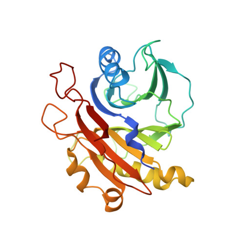
2F1N - PubMed Abstract:
Cytolethal distending toxin (CDT) induces cell cycle arrest and apoptosis in eukaryotic cells, which are mediated by the DNA-damaging CdtB subunit. Here we report the first x-ray structure of an isolated CdtB subunit (Escherichia coli-II CdtB, EcCdtB). In conjunction with previous structural and biochemical observations, active site structural comparisons between free and holotoxin-assembled CdtBs suggested that CDT intoxication is contingent upon holotoxin disassembly. Solution NMR structural and 15N relaxation studies of free EcCdtB revealed disorder in the interface with the CdtA and CdtC subunits (residues Gly233-Asp242). Residues Leu186-Thr209 of EcCdtB, which encompasses tandem arginine residues essential for nuclear translocation and intoxication, were also disordered in solution. In stark contrast, nearly identical well defined alpha-helix and beta-strand secondary structures were observed in this region of the free and holotoxin CdtB crystallographic models, suggesting that distinct changes in structural ordering characterize subunit disassembly and nuclear localization factor binding functions.
Organizational Affiliation:
Division of Cell Biology and Biophysics, School of Biological Sciences, University of Missouri, Kansas City, Missouri 64110-2499, USA.