


Conformational flexibility in the PDZ domain of Dishevelled induced by target binding
Friedland, N., Hung, L.-W., Cheyette, B., Miller, J.R., Moon, R.T., Earnest, T.N.To be published.
Experimental Data Snapshot
wwPDB Validation 3D Report Full Report

Entity ID: 1 | |||||
|---|---|---|---|---|---|
| Molecule | Chains | Sequence Length | Organism | Details | Image |
| Segment polarity protein dishevelled homolog DVL-2 | 98 | Xenopus laevis | Mutation(s): 4 Gene Names: dvl2 | 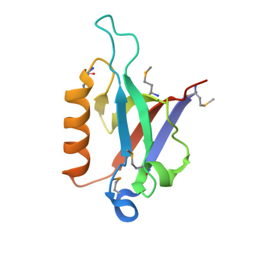 | |
UniProt | |||||
Find proteins for P51142 (Xenopus laevis) Explore P51142 Go to UniProtKB: P51142 | |||||
Entity Groups | |||||
| Sequence Clusters | 30% Identity50% Identity70% Identity90% Identity95% Identity100% Identity | ||||
| UniProt Group | P51142 | ||||
Sequence AnnotationsExpand | |||||
| |||||
| Ligands 2 Unique | |||||
|---|---|---|---|---|---|
| ID | Chains | Name / Formula / InChI Key | 2D Diagram | 3D Interactions | |
| SO4 Query on SO4 | F [auth D] | SULFATE ION O4 S QAOWNCQODCNURD-UHFFFAOYSA-L |  | ||
| CO Query on CO | E [auth B] | COBALT (II) ION Co XLJKHNWPARRRJB-UHFFFAOYSA-N |  | ||
| Modified Residues 1 Unique | |||||
|---|---|---|---|---|---|
| ID | Chains | Type | Formula | 2D Diagram | Parent |
| MSE Query on MSE | A, B, C, D | L-PEPTIDE LINKING | C5 H11 N O2 Se |  | MET |
| Length ( Å ) | Angle ( ˚ ) |
|---|---|
| a = 89.834 | α = 90 |
| b = 89.834 | β = 90 |
| c = 82.471 | γ = 120 |
| Software Name | Purpose |
|---|---|
| REFMAC | refinement |
| Adxv | data processing |
| HKL-2000 | data scaling |
| SOLVE | phasing |
RCSB PDB (citation) is hosted by
RCSB PDB is a member of the

