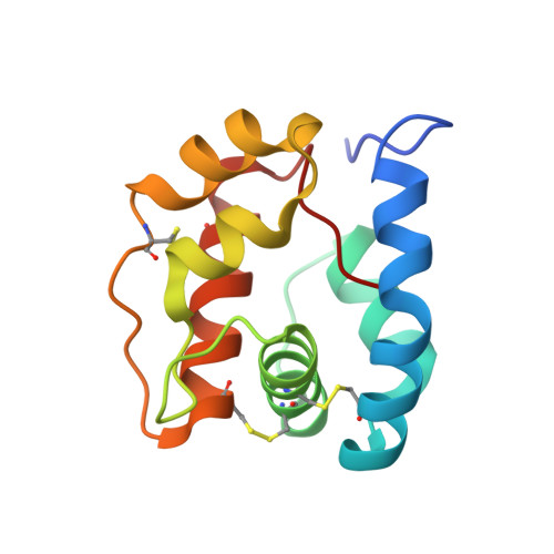
2ERB

AgamOBP1, and odorant binding protein from Anopheles gambiae complexed with PEG


Experimental Data Snapshot

  • Method: X-RAY DIFFRACTION
  • Resolution: 1.50 Å
  • R-Value Free: 0.204 
  • R-Value Work: 0.172 
  • R-Value Observed: 0.172 

wwPDB Validation   3D Report Full Report


This is version 1.3 of the entry. See complete history


Literature

The crystal structure of an odorant binding protein from Anopheles gambiae: Evidence for a common ligand release mechanism.

Wogulis, M.Morgan, T.Ishida, Y.Leal, W.S.Wilson, D.K.

(2006) Biochem Biophys Res Commun 339: 157-164

  • DOI: https://doi.org/10.1016/j.bbrc.2005.10.191
  • Primary Citation of Related Structures:  
    2ERB

  • PubMed Abstract: 

    The Anopheles gambiae mosquito is the main vector of malaria transmission in sub-Saharan Africa. We present here a 1.5A crystal structure of AgamOBP1, an odorant binding protein (OBP) from the A. gambiae mosquito. The protein crystallized as a dimer with a unique binding pocket consisting of a continuous tunnel running through both subunits of the dimer and occupied by a PEG molecule. We demonstrate that AgamOBP1 undergoes a pH dependent conformational change that is associated with reduced ligand binding. A predominance of acid-labile hydrogen bonds involving the C-terminal loop suggests a mechanism in which a drop in pH causes C-terminal loop to open, leaving the binding tunnel solvent exposed, thereby lowering binding affinity for ligand. Because proteins from two distantly related insects also undergo a pH dependent conformational change involving the C-terminus that is associated with reduced ligand affinity, our results suggest a common mechanism for OBP activity.


  • Organizational Affiliation

    Section of Molecular and Cellular Biology, University of California, Davis, CA 95616, USA.


Macromolecules
Find similar proteins by:  (by identity cutoff)  |  3D Structure
Entity ID: 1
MoleculeChains Sequence LengthOrganismDetailsImage
odorant binding protein
A, B
125Anopheles gambiaeMutation(s): 0 
Entity Groups  
Sequence Clusters30% Identity50% Identity70% Identity90% Identity95% Identity100% Identity
Sequence Annotations
Expand
  • Reference Sequence
Small Molecules
Ligands 2 Unique
IDChains Name / Formula / InChI Key2D Diagram3D Interactions
PEU
Query on PEU

Download Ideal Coordinates CCD File 
D [auth B]2,5,8,11,14,17,20,23,26,29,32,35,38,41,44,47,50,53,56,59,62,65,68,71,74,77,80-HEPTACOSAOXADOOCTACONTAN-82-OL
C55 H112 O28
ISGUIIHZEJGUGQ-UHFFFAOYSA-N
MG
Query on MG

Download Ideal Coordinates CCD File 
C [auth A]MAGNESIUM ION
Mg
JLVVSXFLKOJNIY-UHFFFAOYSA-N
Experimental Data & Validation

Experimental Data

  • Method: X-RAY DIFFRACTION
  • Resolution: 1.50 Å
  • R-Value Free: 0.204 
  • R-Value Work: 0.172 
  • R-Value Observed: 0.172 
  • Space Group: P 1 21 1
Unit Cell:
Length ( Å )Angle ( ˚ )
a = 30.1α = 90
b = 68.21β = 99.77
c = 61.48γ = 90
Software Package:
Software NamePurpose
CNSrefinement
MOSFLMdata reduction
CCP4data scaling
CNSphasing

Structure Validation

View Full Validation Report


Entry History 

Deposition Data

Revision History  (Full details and data files)

  • Version 1.0: 2005-12-13
    Type: Initial release
  • Version 1.1: 2008-05-01
    Changes: Version format compliance
  • Version 1.2: 2011-07-13
    Changes: Version format compliance
  • Version 1.3: 2024-04-03
    Changes: Data collection, Database references, Derived calculations, Refinement description