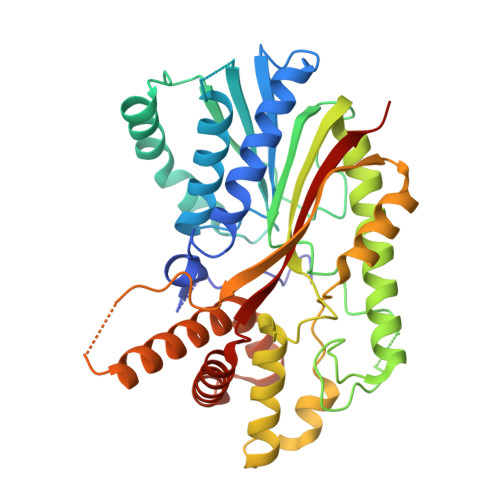
The structure of two N-methyltransferases from the caffeine biosynthetic pathway
McCarthy, A.A., McCarthy, J.G.(2007) Plant Physiol 144: 879-889
- PubMed: 17434991
- DOI: https://doi.org/10.1104/pp.106.094854
- Primary Citation of Related Structures:
2EFJ, 2EG5 - PubMed Abstract:
Caffeine (1,3,7-trimethylxanthine) is a secondary metabolite produced by certain plant species and an important component of coffee (Coffea arabica and Coffea canephora) and tea (Camellia sinensis). Here we describe the structures of two S-adenosyl-l-methionine-dependent N-methyltransferases that mediate caffeine biosynthesis in C. canephora 'robusta', xanthosine (XR) methyltransferase (XMT), and 1,7-dimethylxanthine methyltransferase (DXMT). Both were cocrystallized with the demethylated cofactor, S-adenosyl-L-cysteine, and substrate, either xanthosine or theobromine. Our structures reveal several elements that appear critical for substrate selectivity. Serine-316 in XMT appears central to the recognition of XR. Likewise, a change from glutamine-161 in XMT to histidine-160 in DXMT is likely to have catalytic consequences. A phenylalanine-266 to isoleucine-266 change in DXMT is also likely to be crucial for the discrimination between mono and dimethyl transferases in coffee. These key residues are probably functionally important and will guide future studies with implications for the biosynthesis of caffeine and its derivatives in plants.
Organizational Affiliation:
European Molecular Biology Laboratory, Grenoble 38042, France. andrewmc@embl.fr