


Structure of GK0241 protein from Geobacillus kaustophilus
Lokanath, N.K., Pampa, K.J., Kamiya, N., Kunishima, N.To be published.
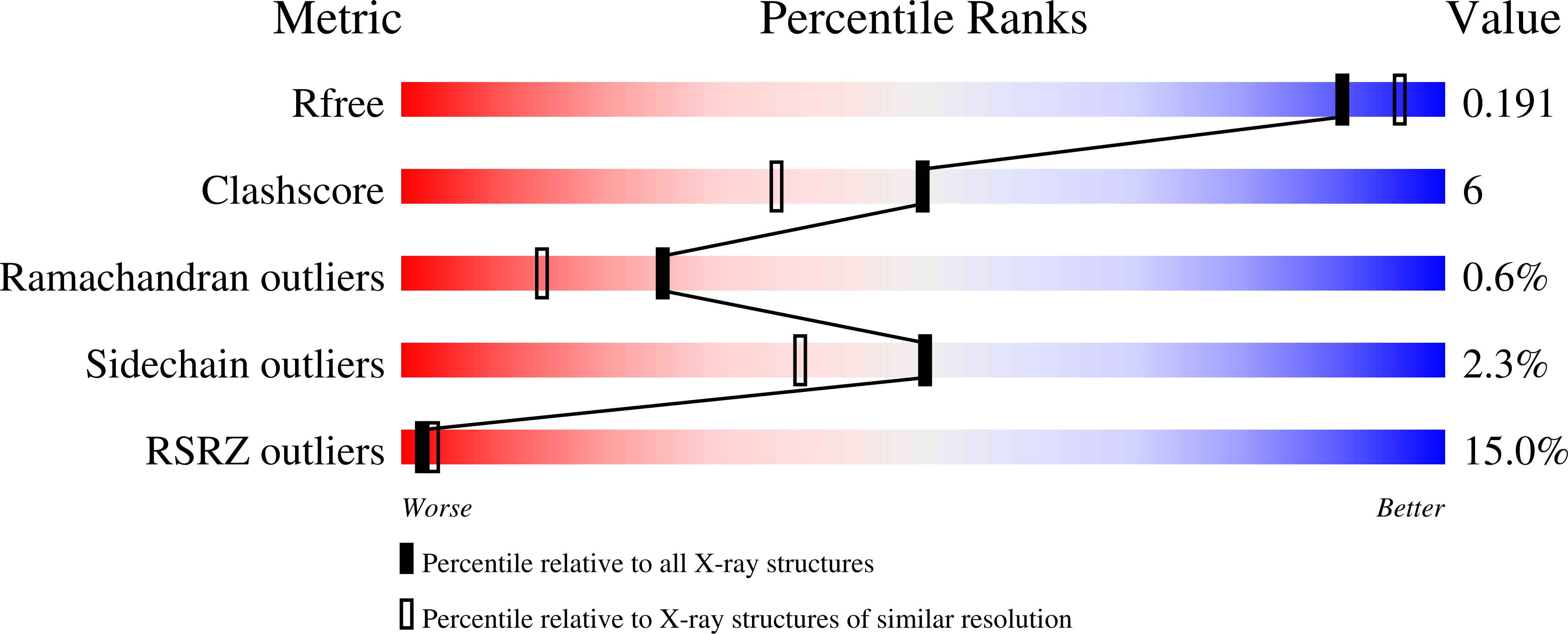
Experimental Data Snapshot
Starting Model: experimental
View more details
wwPDB Validation 3D Report Full Report
Entity ID: 1 | |||||
|---|---|---|---|---|---|
| Molecule | Chains | Sequence Length | Organism | Details | Image |
| Molybdopterin biosynthesis | 162 | Geobacillus kaustophilus HTA426 | Mutation(s): 0 |  | |
UniProt | |||||
Find proteins for Q5L3F4 (Geobacillus kaustophilus (strain HTA426)) Explore Q5L3F4 Go to UniProtKB: Q5L3F4 | |||||
Entity Groups | |||||
| Sequence Clusters | 30% Identity50% Identity70% Identity90% Identity95% Identity100% Identity | ||||
| UniProt Group | Q5L3F4 | ||||
Sequence AnnotationsExpand | |||||
| |||||
| Length ( Å ) | Angle ( ˚ ) |
|---|---|
| a = 105.24 | α = 90 |
| b = 105.24 | β = 90 |
| c = 105.24 | γ = 90 |
| Software Name | Purpose |
|---|---|
| CNS | refinement |
| HKL-2000 | data collection |
| HKL-2000 | data reduction |
| SCALEPACK | data scaling |
| PHASER | phasing |
RCSB PDB (citation) is hosted by
RCSB PDB is a member of the

