2DUV

Structure of CDK2 with a 3-hydroxychromones


Experimental Data Snapshot

  • Method: X-RAY DIFFRACTION
  • Resolution: 2.20 Å
  • R-Value Free: 0.310 
  • R-Value Work: 0.264 
  • R-Value Observed: 0.264 

wwPDB Validation   3D Report Full Report


This is version 1.3 of the entry. See complete history


Literature

3-Hydroxychromones as cyclin-dependent kinase inhibitors: synthesis and biological evaluation.

Lee, J.Park, T.Jeong, S.Kim, K.H.Hong, C.

(2007) Bioorg Med Chem Lett 17: 1284-1287

  • DOI: https://doi.org/10.1016/j.bmcl.2006.12.011
  • Primary Citation of Related Structures:  
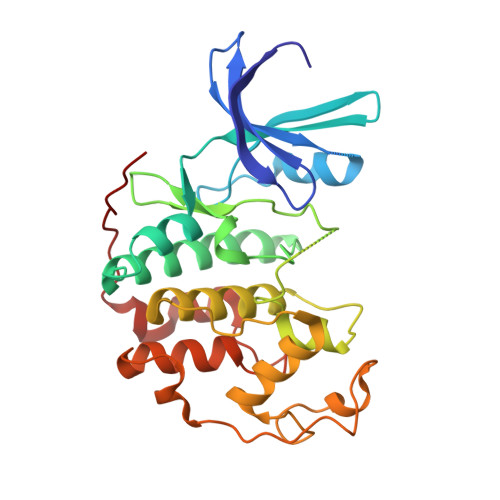
    2DUV

  • PubMed Abstract: 

    A novel series of 3-hydroxychromones were prepared and found to be CDK inhibitors. Isothiazolidine 1,1-dioxide analogues showed potent CDK1 and CDK2 inhibitory activities and inhibited proliferation of EJ, HCT116, SW620, and MDAMB468 cancer cells.


  • Organizational Affiliation

    Department of Chemistry, Keimyung University, 1000 Sindang-Dong, Dalseo-Gu, Daegu 704-701, South Korea. jinho@kmu.ac.kr


Macromolecules
Find similar proteins by:  (by identity cutoff)  |  3D Structure
Entity ID: 1
MoleculeChains Sequence LengthOrganismDetailsImage
Cell division protein kinase 2298Homo sapiensMutation(s): 0 
EC: 2.7.11.22
UniProt & NIH Common Fund Data Resources
Find proteins for P24941 (Homo sapiens)
Explore P24941 
Go to UniProtKB:  P24941
PHAROS:  P24941
GTEx:  ENSG00000123374 
Entity Groups  
Sequence Clusters30% Identity50% Identity70% Identity90% Identity95% Identity100% Identity
UniProt GroupP24941
Sequence Annotations
Expand
  • Reference Sequence
Small Molecules
Ligands 1 Unique
IDChains Name / Formula / InChI Key2D Diagram3D Interactions
371
Query on 371

Download Ideal Coordinates CCD File 
B [auth A]2-(3,4-DIHYDROXYPHENYL)-8-(1,1-DIOXIDOISOTHIAZOLIDIN-2-YL)-3-HYDROXY-6-METHYL-4H-CHROMEN-4-ONE
C19 H17 N O7 S
CEXBEGBNDJVZPK-UHFFFAOYSA-N
Binding Affinity Annotations 
IDSourceBinding Affinity
371 Binding MOAD:  2DUV IC50: 87 (nM) from 1 assay(s)
BindingDB:  2DUV IC50: 87 (nM) from 1 assay(s)
PDBBind:  2DUV IC50: 87 (nM) from 1 assay(s)
Experimental Data & Validation

Experimental Data

  • Method: X-RAY DIFFRACTION
  • Resolution: 2.20 Å
  • R-Value Free: 0.310 
  • R-Value Work: 0.264 
  • R-Value Observed: 0.264 
  • Space Group: P 21 21 21
Unit Cell:
Length ( Å )Angle ( ˚ )
a = 53.629α = 90
b = 71.389β = 90
c = 72.061γ = 90
Software Package:
Software NamePurpose
AMoREphasing
CNSrefinement
DENZOdata reduction
SCALEPACKdata scaling

Structure Validation

View Full Validation Report


Entry History 

Deposition Data

Revision History  (Full details and data files)

  • Version 1.0: 2007-01-27
    Type: Initial release
  • Version 1.1: 2008-04-30
    Changes: Version format compliance
  • Version 1.2: 2011-07-13
    Changes: Version format compliance
  • Version 1.3: 2024-03-13
    Changes: Data collection, Database references, Derived calculations