2DUG

crystal structure of a green fluorescent protein S65T/H148N at pH 5


Experimental Data Snapshot

  • Method: X-RAY DIFFRACTION
  • Resolution: 1.40 Å
  • R-Value Free: 0.241 
  • R-Value Work: 0.177 
  • R-Value Observed: 0.179 

wwPDB Validation   3D Report Full Report


This is version 1.3 of the entry. See complete history


Literature

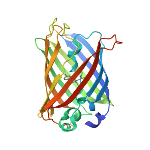
Ultrafast excited-state dynamics in the green fluorescent protein variant S65T/H148D. 1. Mutagenesis and structural studies.

Shu, X.Kallio, K.Shi, X.Abbyad, P.Kanchanawong, P.Childs, W.Boxer, S.G.Remington, S.J.

(2007) Biochemistry 46: 12005-12013

  • DOI: https://doi.org/10.1021/bi7009037
  • Primary Citation of Related Structures:  
    2DUE, 2DUF, 2DUG, 2DUH, 2DUI

  • PubMed Abstract: 

    Wild type green fluorescent protein (wt-GFP) and the variant S65T/H148D each exhibit two absorption bands, A and B, which are associated with the protonated and deprotonated chromophores, respectively. Excitation of either band leads to green emission. In wt-GFP, excitation of band A ( approximately 395 nm) leads to green emission with a rise time of 10-15 ps, due to excited-state proton transfer (ESPT) from the chromophore hydroxyl group to an acceptor. This process produces an anionic excited-state intermediate I* that subsequently emits a green photon. In the variant S65T/H148D, the A band absorbance maximum is red-shifted to approximately 415 nm, and as detailed in the accompanying papers, when the A band is excited, green fluorescence appears with a rise time shorter than the instrument time resolution ( approximately 170 fs). On the basis of the steady-state spectroscopy and high-resolution crystal structures of several variants described herein, it is proposed that in S65T/H148D, the red shift of absorption band A and the ultrafast appearance of green fluorescence upon excitation of band A are due to a very short (


  • Organizational Affiliation

    Institute of Molecular Biology and Department of Physics, University of Oregon, Eugene, Oregon 97403-1229, USA.


Macromolecules
Find similar proteins by:  (by identity cutoff)  |  3D Structure
Entity ID: 1
MoleculeChains Sequence LengthOrganismDetailsImage
Green fluorescent protein236Aequorea victoriaMutation(s): 3 
UniProt
Find proteins for P42212 (Aequorea victoria)
Explore P42212 
Go to UniProtKB:  P42212
Entity Groups  
Sequence Clusters30% Identity50% Identity70% Identity90% Identity95% Identity100% Identity
UniProt GroupP42212
Sequence Annotations
Expand
  • Reference Sequence
Small Molecules
Modified Residues  1 Unique
IDChains TypeFormula2D DiagramParent
C12
Query on C12
A
L-PEPTIDE LINKINGC15 H18 N3 O4THR, TYR, GLY
Experimental Data & Validation

Experimental Data

  • Method: X-RAY DIFFRACTION
  • Resolution: 1.40 Å
  • R-Value Free: 0.241 
  • R-Value Work: 0.177 
  • R-Value Observed: 0.179 
  • Space Group: P 21 21 21
Unit Cell:
Length ( Å )Angle ( ˚ )
a = 51.072α = 90
b = 62.524β = 90
c = 69.072γ = 90
Software Package:
Software NamePurpose
TNTrefinement
d*TREKdata reduction
d*TREKdata scaling
TNTphasing

Structure Validation

View Full Validation Report


Entry History 

Deposition Data

Revision History  (Full details and data files)

  • Version 1.0: 2007-07-10
    Type: Initial release
  • Version 1.1: 2008-04-30
    Changes: Version format compliance
  • Version 1.2: 2011-07-13
    Changes: Version format compliance
  • Version 1.3: 2021-11-10
    Changes: Database references, Derived calculations