


The X-ray crystal structure of formaldehyde dismutase at 2.3 A resolution
Hasegawa, T., Yamano, A., Miura, K., Katsube, Y., Yanase, H., Kato, N.(2002) Acta Crystallogr A 58: C102-C102
Experimental Data Snapshot
(2002) Acta Crystallogr A 58: C102-C102
Entity ID: 1 | |||||
|---|---|---|---|---|---|
| Molecule | Chains | Sequence Length | Organism | Details | Image |
| Formaldehyde dismutase | 398 | Pseudomonas putida | Mutation(s): 0 EC: 1.2.99.4 | 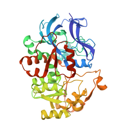 | |
UniProt | |||||
Find proteins for Q52078 (Pseudomonas putida) Explore Q52078 Go to UniProtKB: Q52078 | |||||
Entity Groups | |||||
| Sequence Clusters | 30% Identity50% Identity70% Identity90% Identity95% Identity100% Identity | ||||
| UniProt Group | Q52078 | ||||
Sequence AnnotationsExpand | |||||
| |||||
| Ligands 2 Unique | |||||
|---|---|---|---|---|---|
| ID | Chains | Name / Formula / InChI Key | 2D Diagram | 3D Interactions | |
| NAD Query on NAD | E [auth A], H [auth B] | NICOTINAMIDE-ADENINE-DINUCLEOTIDE C21 H27 N7 O14 P2 BAWFJGJZGIEFAR-NNYOXOHSSA-N |  | ||
| ZN Query on ZN | C [auth A], D [auth A], F [auth B], G [auth B] | ZINC ION Zn PTFCDOFLOPIGGS-UHFFFAOYSA-N |  | ||
| Length ( Å ) | Angle ( ˚ ) |
|---|---|
| a = 90.229 | α = 90 |
| b = 90.229 | β = 90 |
| c = 226.689 | γ = 90 |
| Software Name | Purpose |
|---|---|
| REFMAC | refinement |
| d*TREK | data scaling |
| MLPHARE | phasing |
RCSB PDB (citation) is hosted by
RCSB PDB is a member of the

