2DFX

Crystal structure of the carboxy terminal domain of colicin E5 complexed with its inhibitor


Experimental Data Snapshot

  • Method: X-RAY DIFFRACTION
  • Resolution: 1.90 Å
  • R-Value Free: 0.198 
  • R-Value Work: 0.179 
  • R-Value Observed: 0.180 

wwPDB Validation   3D Report Full Report


This is version 1.3 of the entry. See complete history


Literature

Structural basis for sequence-dependent recognition of colicin E5 tRNase by mimicking the mRNA-tRNA interaction

Yajima, S.Inoue, S.Ogawa, T.Nonaka, T.Ohsawa, K.Masaki, H.

(2006) Nucleic Acids Res 34: 6074-6082

  • DOI: https://doi.org/10.1093/nar/gkl729
  • Primary Citation of Related Structures:  
    2DFX, 2DJH

  • PubMed Abstract: 

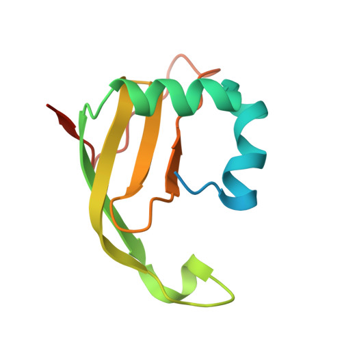
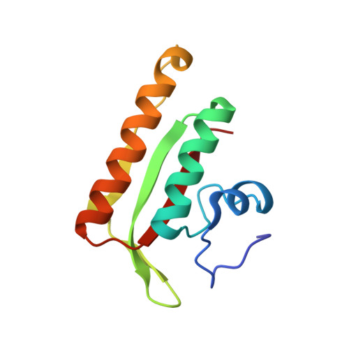
    Colicin E5--a tRNase toxin--specifically cleaves QUN (Q: queuosine) anticodons of the Escherichia coli tRNAs for Tyr, His, Asn and Asp. Here, we report the crystal structure of the C-terminal ribonuclease domain (CRD) of E5 complexed with a substrate analog, namely, dGpdUp, at a resolution of 1.9 A. Thisstructure is the first to reveal the substrate recognition mechanism of sequence-specific ribonucleases. E5-CRD realized the strict recognition for both the guanine and uracil bases of dGpdUp forming Watson-Crick-type hydrogen bonds and ring stacking interactions, thus mimicking the codons of mRNAs to bind to tRNA anticodons. The docking model of E5-CRD with tRNA also suggests its substrate preference for tRNA over ssRNA. In addition, the structure of E5-CRD/dGpdUp along with the mutational analysis suggests that Arg33 may play an important role in the catalytic activity, and Lys25/Lys60 may also be involved without His in E5-CRD. Finally, the comparison of the structures of E5-CRD/dGpdUp and E5-CRD/ImmE5 (an inhibitor protein) complexes suggests that the binding mode of E5-CRD and ImmE5 mimics that of mRNA and tRNA; this may represent the evolutionary pathway of these proteins from the RNA-RNA interaction through the RNA-protein interaction of tRNA/E5-CRD.


  • Organizational Affiliation

    Department of Bioscience, Tokyo University of Agriculture, Sakuragaoka 1-1-1, Setagaya-ku, Tokyo 156-8502, Japan. yshun@nodai.ac.jp


Macromolecules
Find similar proteins by:  (by identity cutoff)  |  3D Structure
Entity ID: 1
MoleculeChains Sequence LengthOrganismDetailsImage
Colicin-E5A [auth E]115Escherichia coliMutation(s): 0 
EC: 3.1
UniProt
Find proteins for P18000 (Escherichia coli)
Explore P18000 
Go to UniProtKB:  P18000
Entity Groups  
Sequence Clusters30% Identity50% Identity70% Identity90% Identity95% Identity100% Identity
UniProt GroupP18000
Sequence Annotations
Expand
  • Reference Sequence
Find similar proteins by:  (by identity cutoff)  |  3D Structure
Entity ID: 2
MoleculeChains Sequence LengthOrganismDetailsImage
Colicin-E5 immunity proteinB [auth I]106Escherichia coliMutation(s): 0 
UniProt
Find proteins for P13476 (Escherichia coli)
Explore P13476 
Go to UniProtKB:  P13476
Entity Groups  
Sequence Clusters30% Identity50% Identity70% Identity90% Identity95% Identity100% Identity
UniProt GroupP13476
Sequence Annotations
Expand
  • Reference Sequence
Experimental Data & Validation

Experimental Data

  • Method: X-RAY DIFFRACTION
  • Resolution: 1.90 Å
  • R-Value Free: 0.198 
  • R-Value Work: 0.179 
  • R-Value Observed: 0.180 
  • Space Group: I 2 2 2
Unit Cell:
Length ( Å )Angle ( ˚ )
a = 63.5α = 90
b = 74.4β = 90
c = 111.2γ = 90
Software Package:
Software NamePurpose
DENZOdata reduction
SCALEPACKdata scaling
MLPHAREphasing
CNSrefinement

Structure Validation

View Full Validation Report


Entry History 

Deposition Data

Revision History  (Full details and data files)

  • Version 1.0: 2007-01-23
    Type: Initial release
  • Version 1.1: 2008-04-30
    Changes: Version format compliance
  • Version 1.2: 2011-07-13
    Changes: Version format compliance
  • Version 1.3: 2024-03-13
    Changes: Data collection, Database references