Structural phase transition of monoclinic crystals of hen egg-white lysozyme
Harata, K., Akiba, T.(2006) Acta Crystallogr D Biol Crystallogr 62: 375-382
- PubMed: 16552138
- DOI: https://doi.org/10.1107/S0907444906001314
- Primary Citation of Related Structures:
2D4I, 2D4J, 2D4K - PubMed Abstract:
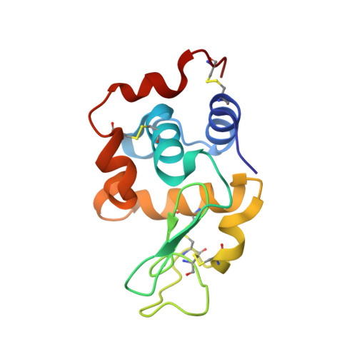
Two monoclinic crystals (space group P2(1)) of hen egg-white lysozyme, a type I crystal grown at room temperature in a D2O solution with pD 4.5 containing 2%(w/v) sodium nitrate and a type II crystal grown at 313 K in a 10%(w/v) sodium chloride solution with pH 7.6, were each transformed into another monoclinic crystal with the same space group by dehydration-induced phase transition. Changes in X-ray diffraction were recorded to monitor the progress of the crystal transformation, which started with the appearance of diffuse streaks. In both crystals, the intensity of h + l odd reflections gradually weakened and finally disappeared on completion of the transformation. X-ray diffraction in the intermediate state indicated the presence of lattices of both the native and transformed crystals. In the native type I crystal, two alternate conformations were observed in the main chain of the region Gly71-Asn74. One conformer bound a sodium ion which was replaced with a water molecule in the other conformer. In the transformed crystal, the sodium ion was removed and the main-chain conformation of this region was converted to that of the water-bound form. The transformed crystal diffracted to a higher resolution than the native crystal, while the peak width of the diffraction spots increased. Analysis of the thermal motion of protein molecules using the TLS model has shown that the enhancement of the diffraction power in the transformed crystal is mainly ascribable to the suppression of rigid-body motion owing to an increase in intermolecular contacts as a result of the loss of bulk solvent.
Organizational Affiliation:
Biological Information Research Center, National Institute of Advanced Industrial Science and Technology (AIST), Central 6, 1-1-1 Higashi, Tsukuba, Ibaraki 305-8566, Japan. k-harata@aist.go.jp