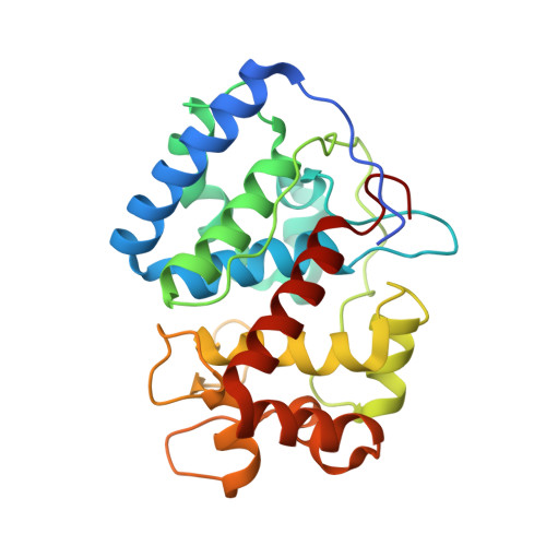
Interaction of Ascorbate Peroxidase with Substrates: A Mechanistic and Structural Analysis.
Macdonald, I.K., Badyal, S.K., Ghamsari, L., Moody, P.C.E., Raven, E.L.(2006) Biochemistry 45: 7808
- PubMed: 16784232
- DOI: https://doi.org/10.1021/bi0606849
- Primary Citation of Related Structures:
2CL4 - PubMed Abstract:
Previous work [Sharp, K. H., et al. (2003) Nat. Struct. Biol. 10, 303-307] has revealed the location of the ascorbate binding site in ascorbate peroxidase and has identified hydrogen-bonding interactions to Arg172, Lys30, and the heme 6-propionate as important in formation of the enzyme-substrate complex. In this work, the individual and collective contributions of these hydrogen bond interactions have been dissected using site-directed mutagenesis, steady-state and pre-steady-state kinetics, X-ray crystallography, and modified substrate analogues. Steady-state and pre-steady-state kinetic data reveal that the hydrogen bonds to Arg172 and the heme 6-propionate play a major part in stabilization of the bound ascorbate but that the interaction with Lys30 plays only a minor role. Binding of aromatic substrates is not affected by substitutions at Arg172/Lys30. Neutralization or removal of electrostatic charge at (Lys30) or adjacent to (Lys31) the ascorbate site does not substantially disrupt the binding interaction. Substrate oxidation and reduction of Compounds I and II is still possible in the absence of Arg172, but at a much reduced level. Crystallographic data (to 1.8 A) for the R172A variant indicate that the molecular structure of the proposed [Sharp, K. H., et al. (2004) Biochemistry 43, 8644-8651] proton transfer pathway from the ascorbate to the heme is conserved, which accounts for the residual activity. The results are discussed in terms of our wider understanding of the structural features that control substrate binding specificity in other peroxidase enzymes.
Organizational Affiliation:
Department of Chemistry, University of Leicester, UK.