2CGK

Crystal Structure of L-rhamnulose kinase from Escherichia coli in an open uncomplexed conformation.


Experimental Data Snapshot

  • Method: X-RAY DIFFRACTION
  • Resolution: 2.46 Å
  • R-Value Free: 0.273 
  • R-Value Work: 0.238 
  • R-Value Observed: 0.238 

wwPDB Validation   3D Report Full Report


This is version 1.4 of the entry. See complete history


Literature

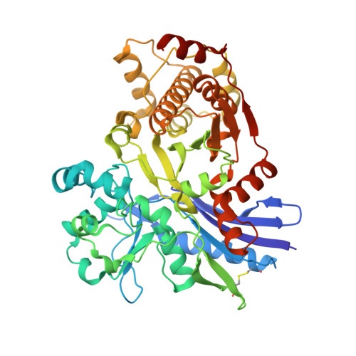
Structure and Reaction Mechanism of L-Rhamnulose Kinase from Escherichia Coli.

Grueninger, D.Schulz, G.E.

(2006) J Mol Biol 359: 787

  • DOI: https://doi.org/10.1016/j.jmb.2006.04.013
  • Primary Citation of Related Structures:  
    2CGJ, 2CGK, 2CGL

  • PubMed Abstract: 

    Bacterial L-rhamnulose kinase participates in the degradation of L-rhamnose, which is ubiquitous and particularly abundant in some plants. The enzyme catalyzes the transfer of the gamma-phosphate group from ATP to the 1-hydroxyl group of L-rhamnulose. We determined the crystal structures of the substrate-free kinase and of a complex between the enzyme, ADP and L-fructose, which besides rhamnulose is also processed. According to its chainfold, the kinase belongs to the hexokinase-hsp70-actin superfamily. The closest structurally known homologue is glycerol kinase. The reported structures reveal a large conformational change on substrate binding as well as the key residues involved in catalysis. The substrates ADP and beta-L-fructose are in an ideal position to define a direct in-line phosphoryl transfer through a bipyramidal pentavalent intermediate. The enzyme contains one disulfide bridge at a position where two homologous glycerol kinases are regulated by phosphorylation and effector binding, respectively, and it has two more pairs of cysteine residues near the surface that are poised for bridging. However, identical catalytic rates were observed for the enzyme in reducing and oxidizing environments, suggesting that regulation by disulfide formation is unlikely.


  • Organizational Affiliation

    Institut für Organische Chemie und Biochemie, Albert-Ludwigs-Universität, Albertstr. 21, 79104 Freiburg im Breisgau, Germany.


Macromolecules
Find similar proteins by:  (by identity cutoff)  |  3D Structure
Entity ID: 1
MoleculeChains Sequence LengthOrganismDetailsImage
L-RHAMNULOSE KINASE
A, B
489Escherichia coli BL21(DE3)Mutation(s): 3 
EC: 2.7.1.5
UniProt
Find proteins for P32171 (Escherichia coli (strain K12))
Explore P32171 
Go to UniProtKB:  P32171
Entity Groups  
Sequence Clusters30% Identity50% Identity70% Identity90% Identity95% Identity100% Identity
UniProt GroupP32171
Sequence Annotations
Expand
  • Reference Sequence
Experimental Data & Validation

Experimental Data

  • Method: X-RAY DIFFRACTION
  • Resolution: 2.46 Å
  • R-Value Free: 0.273 
  • R-Value Work: 0.238 
  • R-Value Observed: 0.238 
  • Space Group: P 1 21 1
Unit Cell:
Length ( Å )Angle ( ˚ )
a = 52.259α = 90
b = 164.933β = 93.45
c = 51.57γ = 90
Software Package:
Software NamePurpose
CNSrefinement
XDSdata reduction
XSCALEdata scaling
CCP4phasing
PHASERphasing
MOLREPphasing

Structure Validation

View Full Validation Report


Entry History 

Deposition Data

Revision History  (Full details and data files)

  • Version 1.0: 2006-05-31
    Type: Initial release
  • Version 1.1: 2011-05-08
    Changes: Version format compliance
  • Version 1.2: 2011-07-13
    Changes: Version format compliance
  • Version 1.3: 2018-10-24
    Changes: Data collection, Source and taxonomy
  • Version 1.4: 2023-12-13
    Changes: Data collection, Database references, Other, Refinement description