2CE2

CRYSTAL STRUCTURE ANALYSIS OF A FLUORESCENT FORM OF H-RAS P21 IN COMPLEX WITH GDP


Experimental Data Snapshot

  • Method: X-RAY DIFFRACTION
  • Resolution: 1.00 Å
  • R-Value Free: 0.163 
  • R-Value Work: 0.144 
  • R-Value Observed: 0.145 

wwPDB Validation   3D Report Full Report


This is version 1.6 of the entry. See complete history


Literature

A Newly Designed Microspectrofluorometer for Kinetic Studies on Protein Crystals in Combination with X-Ray Diffraction

Klink, B.U.Goody, R.S.Scheidig, A.J.

(2006) Biophys J 91: 981

  • DOI: https://doi.org/10.1529/biophysj.105.078931
  • Primary Citation of Related Structures:  
    2CE2, 2CL0, 2CL6, 2CL7, 2CLC, 2CLD, 2EVW

  • PubMed Abstract: 

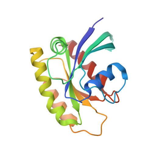
    We present a new design for a fluorescence microspectrophotometer for use in kinetic crystallography in combination with x-ray diffraction experiments. The FLUMIX device (Fluorescence spectroscopy to monitor intermediates in x-ray crystallography) is built for 0 degrees fluorescence detection, which has several advantages in comparison to a conventional fluorometer with 90 degrees design. Due to the reduced spatial requirements and the need for only one objective, the system is highly versatile, easy to handle, and can be used for many different applications. In combination with a conventional stereomicroscope, fluorescence measurements or reaction initiation can be performed directly in a hanging drop crystallization setup. The FLUMIX device can be combined with most x-ray sources, normally without the need of a specialized mechanical support. As a biological model system, we have used H-Ras p21 with an artificially introduced photo-labile GTP precursor (caged GTP) and a covalently attached fluorophore (IANBD amide). Using the FLUMIX system, detailed information about the state of photolyzed crystals of the modified H-Ras p21 (p21(mod)) could be obtained. Measurements in combination with a synchrotron beamline showed significant fluorescence changes in p21(mod) crystals even within a few seconds of x-ray exposure at 100 K.


  • Organizational Affiliation

    Max-Planck-Institut für Molekulare Physiologie, Abteilung Physikalische Biochemie, D-44225 Dortmund, Germany.


Macromolecules
Find similar proteins by:  (by identity cutoff)  |  3D Structure
Entity ID: 1
MoleculeChains Sequence LengthOrganismDetailsImage
GTPASE HRASA [auth X]166Homo sapiensMutation(s): 2 
UniProt & NIH Common Fund Data Resources
Find proteins for P01112 (Homo sapiens)
Explore P01112 
Go to UniProtKB:  P01112
PHAROS:  P01112
GTEx:  ENSG00000174775 
Entity Groups  
Sequence Clusters30% Identity50% Identity70% Identity90% Identity95% Identity100% Identity
UniProt GroupP01112
Sequence Annotations
Expand
  • Reference Sequence
Small Molecules
Ligands 3 Unique
IDChains Name / Formula / InChI Key2D Diagram3D Interactions
GDP
Query on GDP

Download Ideal Coordinates CCD File 
C [auth X]GUANOSINE-5'-DIPHOSPHATE
C10 H15 N5 O11 P2
QGWNDRXFNXRZMB-UUOKFMHZSA-N
XY2
Query on XY2

Download Ideal Coordinates CCD File 
D [auth X]N,N'-DIMETHYL-N-(ACETYL)-N'-(7-NITROBENZ-2-OXA-1,3-DIAZOL-4-YL)ETHYLENEDIAMINE
C12 H15 N5 O4
YSVJZZZGPPWEIC-UHFFFAOYSA-N
MG
Query on MG

Download Ideal Coordinates CCD File 
B [auth X]MAGNESIUM ION
Mg
JLVVSXFLKOJNIY-UHFFFAOYSA-N
Experimental Data & Validation

Experimental Data

  • Method: X-RAY DIFFRACTION
  • Resolution: 1.00 Å
  • R-Value Free: 0.163 
  • R-Value Work: 0.144 
  • R-Value Observed: 0.145 
  • Space Group: C 2 2 21
Unit Cell:
Length ( Å )Angle ( ˚ )
a = 49α = 90
b = 53.9β = 90
c = 116.2γ = 90
Software Package:
Software NamePurpose
REFMACrefinement
XDSdata reduction
XSCALEdata scaling
MOLREPphasing

Structure Validation

View Full Validation Report


Entry History 

Deposition Data

Revision History  (Full details and data files)

  • Version 1.0: 2006-08-23
    Type: Initial release
  • Version 1.1: 2011-07-13
    Changes: Advisory, Version format compliance
  • Version 1.2: 2015-04-22
    Changes: Atomic model, Derived calculations, Non-polymer description, Other, Refinement description
  • Version 1.3: 2017-06-28
    Changes: Database references
  • Version 1.4: 2019-01-30
    Changes: Data collection, Derived calculations, Experimental preparation
  • Version 1.5: 2019-02-06
    Changes: Data collection, Experimental preparation
  • Version 1.6: 2023-12-13
    Changes: Data collection, Database references, Derived calculations, Other, Refinement description