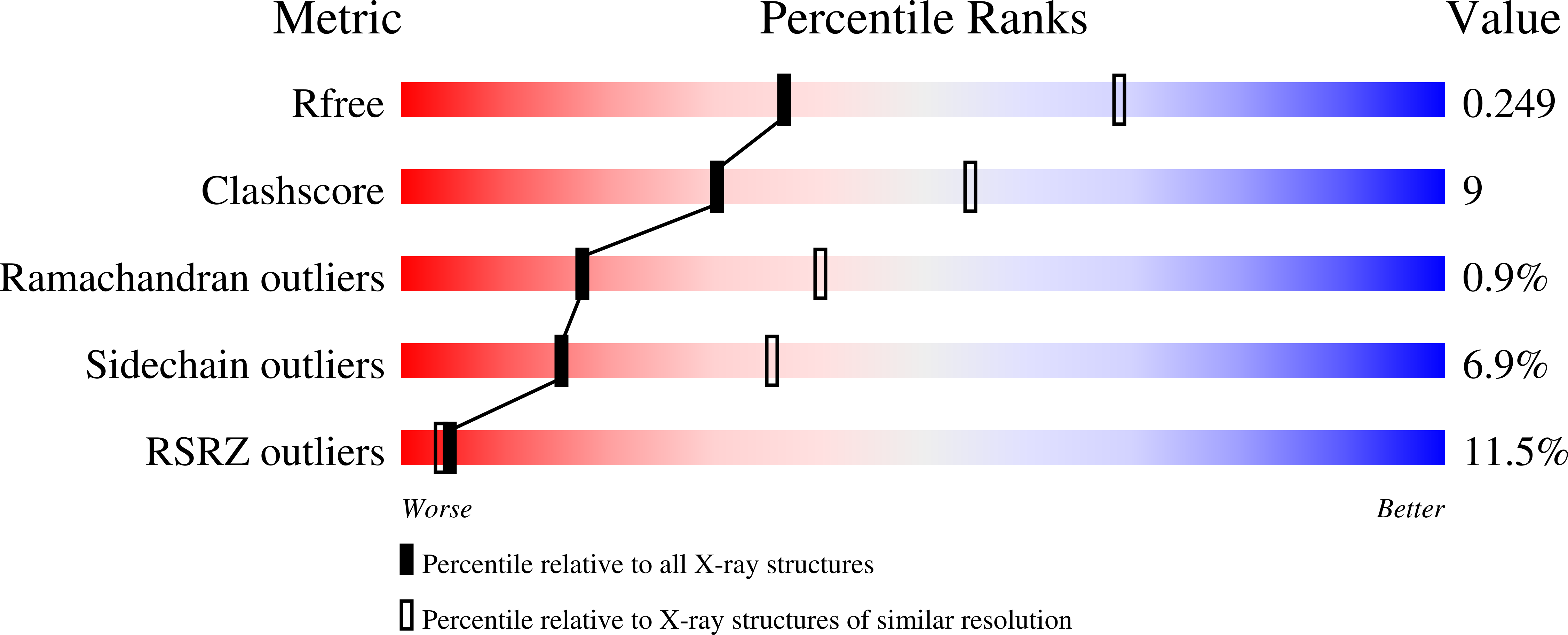
2CDM

The structure of TrwC complexed with a 27-mer DNA comprising the recognition hairpin and the cleavage site


Experimental Data Snapshot

  • Method: X-RAY DIFFRACTION
  • Resolution: 2.70 Å
  • R-Value Free: 0.247 
  • R-Value Work: 0.205 
  • R-Value Observed: 0.207 

wwPDB Validation   3D Report Full Report


This is version 1.4 of the entry. See complete history


Literature

Unveiling the Molecular Mechanism of a Conjugative Relaxase: The Structure of Trwc Complexed with a 27-mer DNA Comprising the Recognition Hairpin and the Cleavage Site.

Boer, R.Russi, S.Guasch, A.Lucas, M.Blanco, A.G.Perez-Luque, R.Coll, M.De La Cruz, F.

(2006) J Mol Biol 358: 857

  • DOI: https://doi.org/10.1016/j.jmb.2006.02.018
  • Primary Citation of Related Structures:  
    1S6M, 1ZM5, 2CDM

  • PubMed Abstract: 

    TrwC is a DNA strand transferase that catalyzes the initial and final stages of conjugative DNA transfer. We have solved the crystal structure of the N-terminal relaxase domain of TrwC in complex with a 27 base-long DNA oligonucleotide that contains both the recognition hairpin and the scissile phosphate. In addition, a series of ternary structures of protein-DNA complexes with different divalent cations at the active site have been solved. Systematic anomalous difference analysis allowed us to determine unambiguously the nature of the metal bound. Zn2+, Ni2+ and Cu2+ were found to bind the histidine-triad metal binding site. Comparison of the structures of the different complexes suggests two pathways for the DNA to exit the active pocket. They are probably used at different steps of the conjugative DNA-processing reaction. The structural information allows us to propose (i) an enzyme mechanism where the scissile phosphate is polarized by the metal ion facilitating the nucleophilic attack of the catalytic tyrosine, and (ii) a probable sequence of events during conjugative DNA processing that explains the biological function of the relaxase.


  • Organizational Affiliation

    Institut de Biologia Molecular de Barcelona (CSIC) and Institut de Recerca Biomèdica, Parc Científic de Barcelona, Josep Samitier 1-5, 08028 Barcelona, Spain.


Macromolecules

Find similar proteins by:  (by identity cutoff)  |  3D Structure
Entity ID: 1
MoleculeChains Sequence LengthOrganismDetailsImage
TRWC
A, C
293Escherichia coliMutation(s): 1 
UniProt
Find proteins for Q47673 (Escherichia coli)
Explore Q47673 
Go to UniProtKB:  Q47673
Entity Groups  
Sequence Clusters30% Identity50% Identity70% Identity90% Identity95% Identity100% Identity
UniProt GroupQ47673
Sequence Annotations
Expand
  • Reference Sequence
Find similar nucleic acids by:  (by identity cutoff)  |  3D Structure
Entity ID: 2
MoleculeChains LengthOrganismImage
5'-D(*GP*CP*GP*CP*AP*CP*CP*GP*AP*AP *AP*GP*GP*TP*GP*CP*GP*TP*AP*TP*TP*GP*TP*CP*TP*AP*T)-3'
B, D
27Escherichia coli
Sequence Annotations
Expand
  • Reference Sequence
Small Molecules
Modified Residues  1 Unique
IDChains TypeFormula2D DiagramParent
MSE
Query on MSE
A, C
L-PEPTIDE LINKINGC5 H11 N O2 SeMET
Experimental Data & Validation

Experimental Data

  • Method: X-RAY DIFFRACTION
  • Resolution: 2.70 Å
  • R-Value Free: 0.247 
  • R-Value Work: 0.205 
  • R-Value Observed: 0.207 
  • Space Group: P 65
Unit Cell:
Length ( Å )Angle ( ˚ )
a = 148.383α = 90
b = 148.383β = 90
c = 79.505γ = 120
Software Package:
Software NamePurpose
REFMACrefinement
MOSFLMdata reduction
SCALAdata scaling

Structure Validation

View Full Validation Report


Entry History 

Deposition Data

Revision History  (Full details and data files)

  • Version 1.0: 2006-07-31
    Type: Initial release
  • Version 1.1: 2011-05-08
    Changes: Version format compliance
  • Version 1.2: 2011-07-13
    Changes: Version format compliance
  • Version 1.3: 2018-01-24
    Changes: Source and taxonomy
  • Version 1.4: 2023-12-13
    Changes: Data collection, Database references, Derived calculations, Other, Refinement description