Structure and Activity of Two Metal-Ion Dependent Acetyl Xylan Esterases Involved in Plant Cell Wall Degradation Reveals a Close Similarity to Peptidoglycan Deacetylases
Taylor, E.J., Gloster, T.M., Turkenburg, J.P., Vincent, F., Brzozowski, A.M., Dupont, C., Shareck, F., Centeno, M.S.J., Prates, J.A.M., Puchart, V., Ferreira, L.M.A., Fontes, C.M.G.A., Biely, P., Davies, G.J.(2006) J Biol Chem 281: 10968
- PubMed: 16431911
- DOI: https://doi.org/10.1074/jbc.M513066200
- Primary Citation of Related Structures:
2C71, 2C79, 2CC0 - PubMed Abstract:
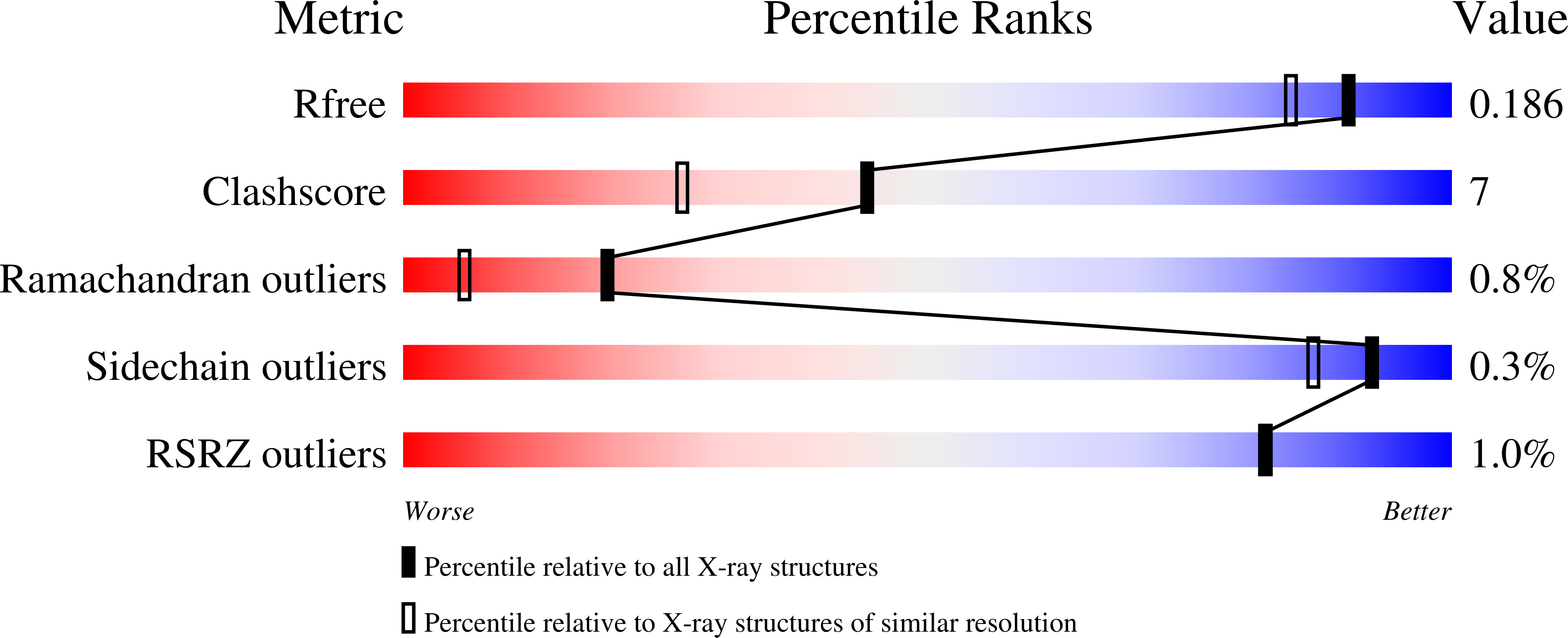
The enzymatic degradation of plant cell wall xylan requires the concerted action of a diverse enzymatic syndicate. Among these enzymes are xylan esterases, which hydrolyze the O-acetyl substituents, primarily at the O-2 position of the xylan backbone. All acetylxylan esterase structures described previously display a alpha/beta hydrolase fold with a "Ser-His-Asp" catalytic triad. Here we report the structures of two distinct acetylxylan esterases, those from Streptomyces lividans and Clostridium thermocellum, in native and complex forms, with x-ray data to between 1.6 and 1.0 A resolution. We show, using a novel linked assay system with PNP-2-O-acetylxyloside and a beta-xylosidase, that the enzymes are sugar-specific and metal ion-dependent and possess a single metal center with a chemical preference for Co2+. Asp and His side chains complete the catalytic machinery. Different metal ion preferences for the two enzymes may reflect the surprising diversity with which the metal ion coordinates residues and ligands in the active center environment of the S. lividans and C. thermocellum enzymes. These "CE4" esterases involved in plant cell wall degradation are shown to be closely related to the de-N-acetylases involved in chitin and peptidoglycan degradation (Blair, D. E., Schuettelkopf, A. W., MacRae, J. I., and Aalten, D. M. (2005) Proc. Natl. Acad. Sci. U. S. A., 102, 15429-15434), which form the NodB deacetylase "superfamily."
Organizational Affiliation:
Structural Biology Laboratory, Department of Chemistry, University of York, Heslington, York YO10 5YW, United Kingdom.


