2BVL

Crystal structure of the catalytic domain of toxin B from Clostridium difficile in complex with UDP, Glc and manganese ion


Experimental Data Snapshot

  • Method: X-RAY DIFFRACTION
  • Resolution: 2.20 Å
  • R-Value Free: 0.244 
  • R-Value Work: 0.197 
  • R-Value Observed: 0.198 

wwPDB Validation   3D Report Full Report


Ligand Structure Quality Assessment 


This is version 1.2 of the entry. See complete history


Literature

Structural Basis for the Function of Clostridium Difficile Toxin B.

Reinert, D.J.Jank, T.Aktories, K.Schulz, G.E.

(2005) J Mol Biol 351: 973

  • DOI: https://doi.org/10.1016/j.jmb.2005.06.071
  • Primary Citation of Related Structures:  
    2BVL, 2BVM

  • PubMed Abstract: 

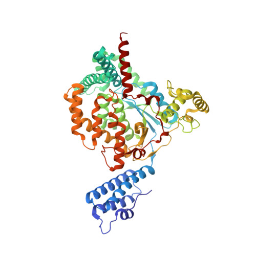
    Toxin B is a member of the family of large clostridial cytotoxins which are of great medical importance. Its catalytic fragment was crystallized in the presence of UDP-glucose and Mn2+. The structure was determined at 2.2 A resolution, showing that toxin B belongs to the glycosyltransferase type A family. However, toxin B contains as many as 309 residues in addition to the common chainfold, which most likely contribute to the target specificity. A superposition with other glycosyltransferases shows the expected positions of the acceptor oxygen atom during glucosyl transfer and indicates further that the reaction proceeds probably along a single-displacement pathway. The C1'' donor carbon atom position is defined by the bound UDP and glucose. It assigns the surface area of toxin B that forms the interface to the target protein during the modifying reaction. A docking attempt brought the known acceptor atom, Thr37 O(gamma1) of the switch I region of the RhoA:GDP target structure, near the expected position. The relative orientation of the two proteins was consistent with both being attached to a membrane. Sequence comparisons between toxin B variants revealed that the highest exchange rate occurs around the active center at the putative docking interface, presumably due to a continuous hit-and-evasion struggle between Clostridia and their eukaryotic hosts.


  • Organizational Affiliation

    Institut für Organische Chemie und Biochemie, Albert-Ludwigs-Universität, Albertstr. 21, 79104 Freiburg im Breisgau, Germany.


Macromolecules
Find similar proteins by:  (by identity cutoff)  |  3D Structure
Entity ID: 1
MoleculeChains Sequence LengthOrganismDetailsImage
TOXIN B543Clostridioides difficileMutation(s): 3 
EC: 2.4.1
UniProt
Find proteins for P18177 (Clostridioides difficile)
Explore P18177 
Go to UniProtKB:  P18177
Entity Groups  
Sequence Clusters30% Identity50% Identity70% Identity90% Identity95% Identity100% Identity
UniProt GroupP18177
Sequence Annotations
Expand
  • Reference Sequence
Small Molecules
Ligands 5 Unique
IDChains Name / Formula / InChI Key2D Diagram3D Interactions
TBR
Query on TBR

Download Ideal Coordinates CCD File 
F [auth A],
G [auth A],
H [auth A],
I [auth A]
HEXATANTALUM DODECABROMIDE
Br12 Ta6
YWYIQTPPCOBSGN-UHFFFAOYSA-M
UDP
Query on UDP

Download Ideal Coordinates CCD File 
C [auth A]URIDINE-5'-DIPHOSPHATE
C9 H14 N2 O12 P2
XCCTYIAWTASOJW-XVFCMESISA-N
GLC
Query on GLC

Download Ideal Coordinates CCD File 
D [auth A]alpha-D-glucopyranose
C6 H12 O6
WQZGKKKJIJFFOK-DVKNGEFBSA-N
SO4
Query on SO4

Download Ideal Coordinates CCD File 
E [auth A]SULFATE ION
O4 S
QAOWNCQODCNURD-UHFFFAOYSA-L
MN
Query on MN

Download Ideal Coordinates CCD File 
B [auth A]MANGANESE (II) ION
Mn
WAEMQWOKJMHJLA-UHFFFAOYSA-N
Experimental Data & Validation

Experimental Data

  • Method: X-RAY DIFFRACTION
  • Resolution: 2.20 Å
  • R-Value Free: 0.244 
  • R-Value Work: 0.197 
  • R-Value Observed: 0.198 
  • Space Group: P 21 21 21
Unit Cell:
Length ( Å )Angle ( ˚ )
a = 63.01α = 90
b = 95.797β = 90
c = 112.915γ = 90
Software Package:
Software NamePurpose
REFMACrefinement
XDSdata reduction
XSCALEdata scaling

Structure Validation

View Full Validation Report


Ligand Structure Quality Assessment 


Entry History 

Deposition Data

Revision History  (Full details and data files)

  • Version 1.0: 2005-08-03
    Type: Initial release
  • Version 1.1: 2011-07-13
    Changes: Advisory, Version format compliance
  • Version 1.2: 2020-07-29
    Type: Remediation
    Reason: Carbohydrate remediation
    Changes: Data collection, Derived calculations, Other, Structure summary