2BU3

Acyl-enzyme intermediate between Alr0975 and glutathione at pH 3.4


Experimental Data Snapshot

  • Method: X-RAY DIFFRACTION
  • Resolution: 1.40 Å
  • R-Value Free: 0.188 
  • R-Value Work: 0.174 
  • R-Value Observed: 0.174 

wwPDB Validation   3D Report Full Report


Ligand Structure Quality Assessment 


This is version 1.2 of the entry. See complete history


Literature

A Papain-Like Enzyme at Work: Native and Acyl- Enzyme Intermediate Structures in Phytochelatin Synthesis.

Vivares, D.Arnoux, P.Pignol, D.

(2005) Proc Natl Acad Sci U S A 102: 18848

  • DOI: https://doi.org/10.1073/pnas.0505833102
  • Primary Citation of Related Structures:  
    2BTW, 2BU3

  • PubMed Abstract: 

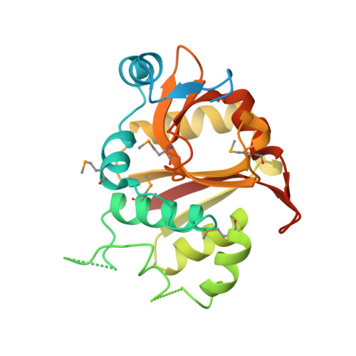
    Phytochelatin synthase (PCS) is a key enzyme for heavy-metal detoxification in plants. PCS catalyzes the production of glutathione (GSH)-derived peptides (called phytochelatins or PCs) that bind heavy-metal ions before vacuolar sequestration. The enzyme can also hydrolyze GSH and GS-conjugated xenobiotics. In the cyanobacterium Nostoc, the enzyme (NsPCS) contains only the catalytic domain of the eukaryotic synthase and can act as a GSH hydrolase and weakly as a peptide ligase. The crystal structure of NsPCS in its native form solved at a 2.0-A resolution shows that NsPCS is a dimer that belongs to the papain superfamily of cysteine proteases, with a conserved catalytic machinery. Moreover, the structure of the protein solved as a complex with GSH at a 1.4-A resolution reveals a gamma-glutamyl cysteine acyl-enzyme intermediate stabilized in a cavity of the protein adjacent to a second putative GSH binding site. GSH hydrolase and PCS activities of the enzyme are discussed in the light of both structures.


  • Organizational Affiliation

    Département d'Ecophysiologie Végétale et de Microbiologie, Direction des Sciences du Vivant, Laboratoire de Bioénergétique Cellulaire, Commissariat á l'Energie Atomique/Cadarache, 13108 St Paul lez Durance Cedex, France.


Macromolecules
Find similar proteins by:  (by identity cutoff)  |  3D Structure
Entity ID: 1
MoleculeChains Sequence LengthOrganismDetailsImage
ALR0975 PROTEIN
A, B
254Anabaena sp.Mutation(s): 0 
EC: 2.3.2.15
UniProt
Find proteins for Q8YY76 (Nostoc sp. (strain PCC 7120 / SAG 25.82 / UTEX 2576))
Explore Q8YY76 
Go to UniProtKB:  Q8YY76
Entity Groups  
Sequence Clusters30% Identity50% Identity70% Identity90% Identity95% Identity100% Identity
UniProt GroupQ8YY76
Sequence Annotations
Expand
  • Reference Sequence
Experimental Data & Validation

Experimental Data

  • Method: X-RAY DIFFRACTION
  • Resolution: 1.40 Å
  • R-Value Free: 0.188 
  • R-Value Work: 0.174 
  • R-Value Observed: 0.174 
  • Space Group: P 1 21 1
Unit Cell:
Length ( Å )Angle ( ˚ )
a = 56.253α = 90
b = 58.259β = 108.87
c = 72.561γ = 90
Software Package:
Software NamePurpose
REFMACrefinement
MOSFLMdata reduction
SCALAdata scaling
MOLREPphasing

Structure Validation

View Full Validation Report


Ligand Structure Quality Assessment 


Entry History 

Deposition Data

Revision History  (Full details and data files)

  • Version 1.0: 2005-12-14
    Type: Initial release
  • Version 1.1: 2011-07-13
    Changes: Advisory, Version format compliance
  • Version 1.2: 2023-12-13
    Changes: Data collection, Database references, Derived calculations, Other, Refinement description