2BRM

Structure-based Design of Novel Chk1 Inhibitors: Insights into Hydrogen Bonding and Protein-Ligand Affinity


Experimental Data Snapshot

  • Method: X-RAY DIFFRACTION
  • Resolution: 2.20 Å
  • R-Value Free: 0.281 
  • R-Value Work: 0.226 
  • R-Value Observed: 0.228 

wwPDB Validation   3D Report Full Report


Ligand Structure Quality Assessment 


This is version 1.3 of the entry. See complete history


Literature

Structure-Based Design of Novel Chk1 Inhibitors: Insights Into Hydrogen Bonding and Protein-Ligand Affinity.

Foloppe, N.Fisher, L.M.Howes, R.Kierstan, P.Potter, A.Robertson, A.G.S.Surgenor, A.E.

(2005) J Med Chem 48: 4332

  • DOI: https://doi.org/10.1021/jm049022c
  • Primary Citation of Related Structures:  
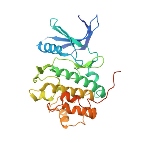
    2BR1, 2BRB, 2BRG, 2BRH, 2BRM, 2BRN, 2BRO

  • PubMed Abstract: 

    We report the discovery, synthesis, and crystallographic binding mode of novel furanopyrimidine and pyrrolopyrimidine inhibitors of the Chk1 kinase, an oncology target. These inhibitors are synthetically tractable and inhibit Chk1 by competing for its ATP site. A chronological account allows an objective comparison of modeled compound docking modes to the subsequently obtained crystal structures. The comparison provides insights regarding the interpretation of modeling results, in relationship to the multiple reasonable docking modes which may be obtained in a kinase-ATP site. The crystal structures were used to guide medicinal chemistry efforts. This led to a thorough characterization of a pair of ligand-protein complexes which differ by a single hydrogen bond. An analysis indicates that this hydrogen bond is expected to contribute a fraction of the 10-fold change in binding affinity, adding a valuable observation to the debate about the energetic role of hydrogen bonding in molecular recognition.


  • Organizational Affiliation

    Vernalis (R&D) Limited, Granta Park, Abington, Cambridge CB1 6GB, UK. n.foloppe@vernalis.com


Macromolecules
Find similar proteins by:  (by identity cutoff)  |  3D Structure
Entity ID: 1
MoleculeChains Sequence LengthOrganismDetailsImage
SERINE/THREONINE-PROTEIN KINASE CHK1297Homo sapiensMutation(s): 0 
EC: 2.7.1.37
UniProt & NIH Common Fund Data Resources
Find proteins for O14757 (Homo sapiens)
Explore O14757 
Go to UniProtKB:  O14757
PHAROS:  O14757
GTEx:  ENSG00000149554 
Entity Groups  
Sequence Clusters30% Identity50% Identity70% Identity90% Identity95% Identity100% Identity
UniProt GroupO14757
Sequence Annotations
Expand
  • Reference Sequence
Small Molecules
Ligands 1 Unique
IDChains Name / Formula / InChI Key2D Diagram3D Interactions
DFZ
Query on DFZ

Download Ideal Coordinates CCD File 
B [auth A]3-AMINO-3-BENZYL-[4.3.0]BICYCLO-1,6-DIAZANONAN-2-ONE
C20 H18 N4 O
MPJYGQHMIFDQIN-UHFFFAOYSA-N
Binding Affinity Annotations 
IDSourceBinding Affinity
DFZ PDBBind:  2BRM Ki: 1300 (nM) from 1 assay(s)
Binding MOAD:  2BRM Ki: 1300 (nM) from 1 assay(s)
BindingDB:  2BRM IC50: 2300 (nM) from 1 assay(s)
Experimental Data & Validation

Experimental Data

  • Method: X-RAY DIFFRACTION
  • Resolution: 2.20 Å
  • R-Value Free: 0.281 
  • R-Value Work: 0.226 
  • R-Value Observed: 0.228 
  • Space Group: P 1 21 1
Unit Cell:
Length ( Å )Angle ( ˚ )
a = 45.116α = 90
b = 65.909β = 101.38
c = 54.582γ = 90
Software Package:
Software NamePurpose
REFMACrefinement
DENZOdata reduction
SCALEPACKdata scaling
AMoREphasing

Structure Validation

View Full Validation Report


Ligand Structure Quality Assessment 


Entry History 

Deposition Data

Revision History  (Full details and data files)

  • Version 1.0: 2005-05-12
    Type: Initial release
  • Version 1.1: 2011-05-08
    Changes: Version format compliance
  • Version 1.2: 2011-07-13
    Changes: Version format compliance
  • Version 1.3: 2023-12-13
    Changes: Data collection, Database references, Other, Refinement description