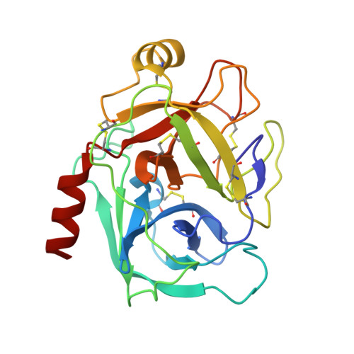
2AYW

Crystal Structure of the complex formed between trypsin and a designed synthetic highly potent inhibitor in the presence of benzamidine at 0.97 A resolution


Experimental Data Snapshot

  • Method: X-RAY DIFFRACTION
  • Resolution: 0.97 Å
  • R-Value Free: 0.157 
  • R-Value Work: 0.134 
  • R-Value Observed: 0.138 

wwPDB Validation   3D Report Full Report


Ligand Structure Quality Assessment 


This is version 1.4 of the entry. See complete history


Literature

Structure of the complex of trypsin with a highly potent synthetic inhibitor at 0.97 A resolution.

Sherawat, M.Kaur, P.Perbandt, M.Betzel, C.Slusarchyk, W.A.Bisacchi, G.S.Chang, C.Jacobson, B.L.Einspahr, H.M.Singh, T.P.

(2007) Acta Crystallogr D Biol Crystallogr 63: 500-507

  • DOI: https://doi.org/10.1107/S090744490700697X
  • Primary Citation of Related Structures:  
    2AYW

  • PubMed Abstract: 

    The structure of the complex formed between bovine beta-trypsin and the highly potent synthetic inhibitor 2-{3'-[5'-methoxy-2'-(N-p-diaminomethylphenyl)amido]-1'-pyrido}-5-(N-2''-t-butylethanol)amidobenzoic acid (C(28)H(32)N(5)O(6)) has been determined at 0.97 A resolution. X-ray intensity data were collected to 0.97 A under cryocooled conditions. The structure was refined anisotropically using REFMAC5 and SHELX-97 to R(cryst) factors of 13.4 and 12.6% and R(free) factors of 15.7 and 16.3%, respectively. Several regions of the main chain and side chains that have not been previously observed were clearly defined in the present structure. H atoms are indicated as significant peaks in an |F(o) - F(c)| difference map, which accounts for an estimated 35% of all H atoms at the 2.5sigma level. The C, N and O atoms are definitively differentiated in the electron-density maps. The amido part of the inhibitor occupies the specificity pocket and the remainder fills the remaining part of the ligand-binding cleft and interacts with the enzyme through an extensive network of hydrogen bonds. The inhibitor distorts the stereochemistry of the catalytic triad, Ser195-His57-Asp102, thereby blocking the proton-relay process of the active site by preventing the formation of the crucial hydrogen bond between His57 N(delta1) and Asp102 O(delta1).


  • Organizational Affiliation

    Department of Biophysics, All India Institute of Medical Sciences, New Delhi 110029, India.


Macromolecules
Find similar proteins by:  (by identity cutoff)  |  3D Structure
Entity ID: 1
MoleculeChains Sequence LengthOrganismDetailsImage
Cationic trypsin223Bos taurusMutation(s): 0 
EC: 3.4.21.4
UniProt
Find proteins for P00760 (Bos taurus)
Explore P00760 
Go to UniProtKB:  P00760
Entity Groups  
Sequence Clusters30% Identity50% Identity70% Identity90% Identity95% Identity100% Identity
UniProt GroupP00760
Sequence Annotations
Expand
  • Reference Sequence
Small Molecules
Ligands 5 Unique
IDChains Name / Formula / InChI Key2D Diagram3D Interactions
ONO
Query on ONO

Download Ideal Coordinates CCD File 
C [auth A],
D [auth A]
2-[2-({[4-(DIAMINOMETHYL)PHENYL]AMINO}CARBONYL)-6-METHOXYPYRIDIN-3-YL]-5-{[(1-FORMYL-2,2-DIMETHYLPROPYL)AMINO]CARBONYL}BENZOIC ACID
C28 H31 N5 O6
XTFRBCPDFSHPBF-OAQYLSRUSA-N
MES
Query on MES

Download Ideal Coordinates CCD File 
F [auth A]2-(N-MORPHOLINO)-ETHANESULFONIC ACID
C6 H13 N O4 S
SXGZJKUKBWWHRA-UHFFFAOYSA-N
BEN
Query on BEN

Download Ideal Coordinates CCD File 
E [auth A]BENZAMIDINE
C7 H8 N2
PXXJHWLDUBFPOL-UHFFFAOYSA-N
GOL
Query on GOL

Download Ideal Coordinates CCD File 
G [auth A]GLYCEROL
C3 H8 O3
PEDCQBHIVMGVHV-UHFFFAOYSA-N
CA
Query on CA

Download Ideal Coordinates CCD File 
B [auth A]CALCIUM ION
Ca
BHPQYMZQTOCNFJ-UHFFFAOYSA-N
Experimental Data & Validation

Experimental Data

  • Method: X-RAY DIFFRACTION
  • Resolution: 0.97 Å
  • R-Value Free: 0.157 
  • R-Value Work: 0.134 
  • R-Value Observed: 0.138 
  • Space Group: P 32 2 1
Unit Cell:
Length ( Å )Angle ( ˚ )
a = 68.55α = 90
b = 68.55β = 90
c = 73.33γ = 120
Software Package:
Software NamePurpose
REFMACrefinement
XDSdata reduction
SCALEPACKdata scaling
AMoREphasing

Structure Validation

View Full Validation Report


Ligand Structure Quality Assessment 


Entry History 

Deposition Data

Revision History  (Full details and data files)

  • Version 1.0: 2006-01-17
    Type: Initial release
  • Version 1.1: 2008-05-01
    Changes: Version format compliance
  • Version 1.2: 2011-07-13
    Changes: Non-polymer description, Version format compliance
  • Version 1.3: 2017-10-11
    Changes: Refinement description
  • Version 1.4: 2023-10-25
    Changes: Data collection, Database references, Derived calculations, Refinement description