Functional and Structural Characterization of Spl Proteases from Staphylococcus aureus
Popowicz, G.M., Dubin, G., Stec-Niemczyk, J., Czarny, A., Dubin, A., Potempa, J., Holak, T.A.(2006) J Mol Biol 358: 270-279
- PubMed: 16516230
- DOI: https://doi.org/10.1016/j.jmb.2006.01.098
- Primary Citation of Related Structures:
2AS9 - PubMed Abstract:
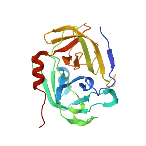
Staphylococcus aureus is the major cause of nosocomial infections world-wide, with increasing prevalence of community-acquired diseases. The recent dramatic increase in multi-antibiotic resistance, including resistance to the last-resort drug, vancomycin, together with the lack of an effective vaccine highlight the need for better understanding of S.aureus pathogenicity. Comparative analysis of available bacterial genomes allows for the identification of previously uncharacterized S.aureus genes with potential roles in pathogenicity. A good example is a cluster of six serine protease-like (spl) genes encompassed in one operon, which encode for putative proteases with similarity to staphylococcal glutamylendopeptidase (V8 protease). Here, we describe an efficient expression system for the production of recombinant SplB and SplC proteases in Escherichia coli, together with structural and functional characterization of the purified enzymes. A unique mechanism of cytoplasm protection against activity of misdirected SplB was uncovered. Apparently, the co-translated signal peptide maintains protease latency until it is cleaved by the signal peptidase during protein secretion. Furthermore, the crystal structure of the SplC protease revealed a fold resembling that of the V8 protease and epidermolytic toxins. Arrangement of the active site cleft and substrate-binding pocket of SplC explains the mechanism of enzyme latency and suggests that some Spl proteases possess restricted substrate specificity similar to that of the V8 protease and epidermolytic toxins.
Organizational Affiliation:
Max-Planck Institute of Biochemistry, Am Klopferspitz 18A, 82-152 Martinsried, Germany.