Functional significance of Glu-77 and Tyr-137 within the active site of isoaspartyl dipeptidase.
Marti-Arbona, R., Thoden, J.B., Holden, H.M., Raushel, F.M.(2005) Bioorg Chem 33: 448-458
- PubMed: 16289685
- DOI: https://doi.org/10.1016/j.bioorg.2005.10.002
- Primary Citation of Related Structures:
2AQO, 2AQV - PubMed Abstract:
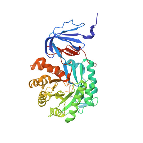
Isoaspartyl dipeptidase (IAD) is a binuclear metalloenzyme and a member of the amidohydrolase superfamily. This enzyme catalyzes the hydrolytic cleavage of beta-aspartyl dipeptides. The pH-rate profiles for the hydrolysis of beta-Asp-Leu indicates that catalysis is dependent on the ionization of two groups; one that ionizes at a pH approximately 6 and the other approximately 9. The group that must be ionized for catalysis is directly dependent on the identity of the metal ion bound to the active site. This result is consistent with the ionization of the hydroxide that bridges the two divalent cations. In addition to the residues that interact directly with the divalent cations there are two other residues that are highly conserved and found within the active site: Glu-77 and Tyr-137. Mutation of Tyr-137 to phenylalanine reduced the rate of catalysis by three orders of magnitude. The three dimensional X-ray structure of the Y137F mutant did not show any significant conformation changes relative to the three dimensional structure of the wild-type enzyme. The positioning of the side-chain phenolic group of Tyr-137 in the active site of IAD is consistent with the stabilization of the tetrahedral adduct concomitant with nucleophilic attack by the hydroxide that bridges the two divalent cations. Mutation of Glu-77 resulted in the reduction of catalytic activity by five orders of magnitude. The three dimensional structure of the E77Q mutant did not show any significant conformational changes in the mutant relative to the three dimensional structure of the wild-type enzyme. The positioning of the side-chain carboxylate of Glu-77 is consistent with the formation of an ion pair interaction with the free alpha-amino group of the substrate.
Organizational Affiliation:
Department of Biochemistry, University of Wisconsin, Madison, WI 53706, USA.