2AGG

succinyl-AAPK-trypsin acyl-enzyme at 1.28 A resolution


Experimental Data Snapshot

  • Method: X-RAY DIFFRACTION
  • Resolution: 1.28 Å
  • R-Value Free: 0.158 
  • R-Value Work: 0.124 
  • R-Value Observed: 0.125 

wwPDB Validation   3D Report Full Report


This is version 1.4 of the entry. See complete history


Literature

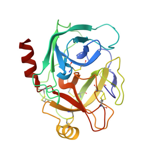
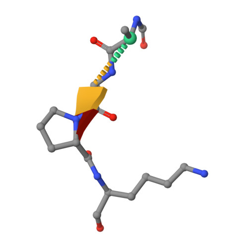
Insights into the serine protease mechanism from atomic resolution structures of trypsin reaction intermediates

Radisky, E.S.Lee, J.M.Lu, C.J.Koshland Jr., D.E.

(2006) Proc Natl Acad Sci U S A 103: 6835-6840

  • DOI: https://doi.org/10.1073/pnas.0601910103
  • Primary Citation of Related Structures:  
    2AGE, 2AGG, 2AGI, 2AH4

  • PubMed Abstract: 

    Atomic resolution structures of trypsin acyl-enzymes and a tetrahedral intermediate analog, along with previously solved structures representing the Michaelis complex, are used to reconstruct events in the catalytic cycle of this classic serine protease. Structural comparisons provide insight into active site adjustments involved in catalysis. Subtle motions of the catalytic serine and histidine residues coordinated with translation of the substrate reaction center are seen to favor the forward progress of the acylation reaction. The structures also clarify the attack trajectory of the hydrolytic water in the deacylation reaction.


  • Organizational Affiliation

    Department of Molecular and Cell Biology, University of California, Berkeley, CA 94720, USA.


Macromolecules
Find similar proteins by:  (by identity cutoff)  |  3D Structure
Entity ID: 1
MoleculeChains Sequence LengthOrganismDetailsImage
beta-trypsinA [auth X]223Bos taurusMutation(s): 0 
EC: 3.4.21.4
UniProt
Find proteins for P00760 (Bos taurus)
Explore P00760 
Go to UniProtKB:  P00760
Entity Groups  
Sequence Clusters30% Identity50% Identity70% Identity90% Identity95% Identity100% Identity
UniProt GroupP00760
Sequence Annotations
Expand
  • Reference Sequence

Find similar proteins by:  Sequence   |   3D Structure  

Entity ID: 2
MoleculeChains Sequence LengthOrganismDetailsImage
succinyl-Ala-Ala-Pro-LysB [auth A]5N/AMutation(s): 0 
Sequence Annotations
Expand
  • Reference Sequence
Biologically Interesting Molecules (External Reference) 1 Unique
Experimental Data & Validation

Experimental Data

  • Method: X-RAY DIFFRACTION
  • Resolution: 1.28 Å
  • R-Value Free: 0.158 
  • R-Value Work: 0.124 
  • R-Value Observed: 0.125 
  • Space Group: P 21 21 21
Unit Cell:
Length ( Å )Angle ( ˚ )
a = 61.388α = 90
b = 63.588β = 90
c = 68.849γ = 90
Software Package:
Software NamePurpose
MOSFLMdata reduction
SCALAdata scaling
EPMRphasing
REFMACrefinement
CCP4data scaling

Structure Validation

View Full Validation Report


Entry History 

Deposition Data

Revision History  (Full details and data files)

  • Version 1.0: 2006-05-16
    Type: Initial release
  • Version 1.1: 2008-04-01
    Changes: Version format compliance
  • Version 1.2: 2011-07-13
    Changes: Atomic model, Database references, Derived calculations, Non-polymer description, Structure summary, Version format compliance
  • Version 1.3: 2012-12-12
    Changes: Other
  • Version 1.4: 2023-08-23
    Changes: Advisory, Data collection, Database references, Derived calculations, Refinement description