2A99

Crystal structure of recombinant chicken sulfite oxidase at resting state


Experimental Data Snapshot

  • Method: X-RAY DIFFRACTION
  • Resolution: 2.20 Å
  • R-Value Free: 0.182 
  • R-Value Work: 0.149 
  • R-Value Observed: 0.150 

wwPDB Validation   3D Report Full Report


Ligand Structure Quality Assessment 


This is version 1.4 of the entry. See complete history


Literature

Structural insights into sulfite oxidase deficiency

Karakas, E.Wilson, H.L.Graf, T.N.Xiang, S.Jaramillo-Busquets, S.Rajagopalan, K.V.Kisker, C.

(2005) J Biol Chem 280: 33506-33515

  • DOI: https://doi.org/10.1074/jbc.M505035200
  • Primary Citation of Related Structures:  
    2A99, 2A9A, 2A9B, 2A9C, 2A9D

  • PubMed Abstract: 

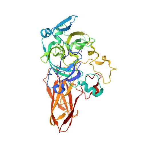
    Sulfite oxidase deficiency is a lethal genetic disease that results from defects either in the genes encoding proteins involved in molybdenum cofactor biosynthesis or in the sulfite oxidase gene itself. Several point mutations in the sulfite oxidase gene have been identified from patients suffering from this disease worldwide. Although detailed biochemical analyses have been carried out on these mutations, no structural data could be obtained because of problems in crystallizing recombinant human and rat sulfite oxidases and the failure to clone the chicken sulfite oxidase gene. We synthesized the gene for chicken sulfite oxidase de novo, working backward from the amino acid sequence of the native chicken liver enzyme by PCR amplification of a series of 72 overlapping primers. The recombinant protein displayed the characteristic absorption spectrum of sulfite oxidase and exhibited steady state and rapid kinetic parameters comparable with those of the tissue-derived enzyme. We solved the crystal structures of the wild type and the sulfite oxidase deficiency-causing R138Q (R160Q in humans) variant of recombinant chicken sulfite oxidase in the resting and sulfate-bound forms. Significant alterations in the substrate-binding pocket were detected in the structure of the mutant, and a comparison between the wild type and mutant protein revealed that the active site residue Arg-450 adopts different conformations in the presence and absence of bound sulfate. The size of the binding pocket is thereby considerably reduced, and its position relative to the cofactor is shifted, causing an increase in the distance of the sulfur atom of the bound sulfate to the molybdenum.


  • Organizational Affiliation

    Department of Pharmacological Sciences, State University of New York, Stony Brook, New York 11794-5115, USA.


Macromolecules
Find similar proteins by:  (by identity cutoff)  |  3D Structure
Entity ID: 1
MoleculeChains Sequence LengthOrganismDetailsImage
Sulfite Oxidase372Gallus gallusMutation(s): 0 
Gene Names: SUOX
EC: 1.8.3.1
UniProt
Find proteins for P07850 (Gallus gallus)
Explore P07850 
Go to UniProtKB:  P07850
Entity Groups  
Sequence Clusters30% Identity50% Identity70% Identity90% Identity95% Identity100% Identity
UniProt GroupP07850
Sequence Annotations
Expand
  • Reference Sequence
Small Molecules
Ligands 4 Unique
IDChains Name / Formula / InChI Key2D Diagram3D Interactions
MTE
Query on MTE

Download Ideal Coordinates CCD File 
C [auth A]PHOSPHONIC ACIDMONO-(2-AMINO-5,6-DIMERCAPTO-4-OXO-3,7,8A,9,10,10A-HEXAHYDRO-4H-8-OXA-1,3,9,10-TETRAAZA-ANTHRACEN-7-YLMETHYL)ESTER
C10 H14 N5 O6 P S2
HPEUEJRPDGMIMY-IFQPEPLCSA-N
MO
Query on MO

Download Ideal Coordinates CCD File 
D [auth A]MOLYBDENUM ATOM
Mo
ZOKXTWBITQBERF-UHFFFAOYSA-N
GOL
Query on GOL

Download Ideal Coordinates CCD File 
E [auth A]GLYCEROL
C3 H8 O3
PEDCQBHIVMGVHV-UHFFFAOYSA-N
CL
Query on CL

Download Ideal Coordinates CCD File 
B [auth A]CHLORIDE ION
Cl
VEXZGXHMUGYJMC-UHFFFAOYSA-M
Experimental Data & Validation

Experimental Data

  • Method: X-RAY DIFFRACTION
  • Resolution: 2.20 Å
  • R-Value Free: 0.182 
  • R-Value Work: 0.149 
  • R-Value Observed: 0.150 
  • Space Group: I 41
Unit Cell:
Length ( Å )Angle ( ˚ )
a = 86.004α = 90
b = 86.004β = 90
c = 154.18γ = 90
Software Package:
Software NamePurpose
HKL-2000data collection
DENZOdata reduction
SCALEPACKdata scaling
MOLREPphasing
REFMACrefinement
HKL-2000data reduction
HKL-2000data scaling

Structure Validation

View Full Validation Report


Ligand Structure Quality Assessment 


Entry History 

Deposition Data

Revision History  (Full details and data files)

  • Version 1.0: 2005-08-02
    Type: Initial release
  • Version 1.1: 2008-04-30
    Changes: Version format compliance
  • Version 1.2: 2011-07-13
    Changes: Advisory, Version format compliance
  • Version 1.3: 2017-10-11
    Changes: Refinement description
  • Version 1.4: 2023-08-23
    Changes: Data collection, Database references, Derived calculations, Refinement description