2A6J

Crystal structure analysis of the anti-arsonate germline antibody 36-65


Experimental Data Snapshot

  • Method: X-RAY DIFFRACTION
  • Resolution: 2.70 Å
  • R-Value Free: 0.265 
  • R-Value Work: 0.225 
  • R-Value Observed: 0.228 

wwPDB Validation   3D Report Full Report


This is version 1.4 of the entry. See complete history


Literature

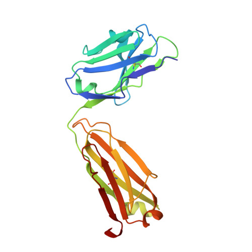
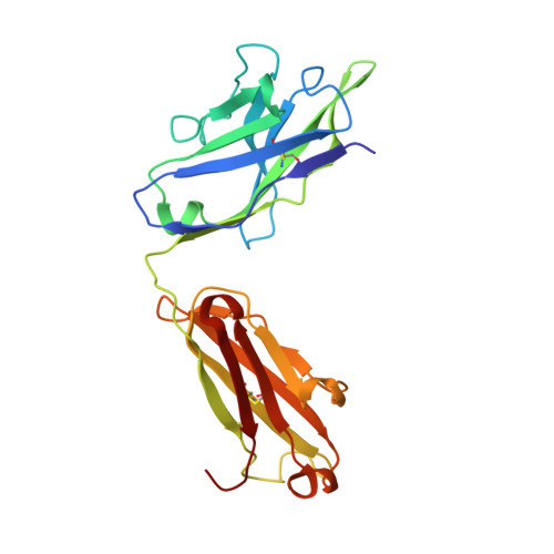
Differential epitope positioning within the germline antibody paratope enhances promiscuity in the primary immune response.

Sethi, D.K.Agarwal, A.Manivel, V.Rao, K.V.Salunke, D.M.

(2006) Immunity 24: 429-438

  • DOI: https://doi.org/10.1016/j.immuni.2006.02.010
  • Primary Citation of Related Structures:  
    2A6D, 2A6I, 2A6J, 2A6K

  • PubMed Abstract: 

    Correlation between the promiscuity of the primary antibody response and conformational flexibility in a germline antibody was addressed by using germline antibody 36-65. Crystallographic analyses of the 36-65 Fab with three independent dodecapeptides provided mechanistic insights into the generation of antibody diversity. While four antigen-free Fab molecules provided a quantitative description of the conformational repertoire of the antibody CDRs, three Fab molecules bound to structurally diverse peptide epitopes exhibited a common paratope conformation. Each peptide revealed spatially different footprints within the antigen-combining site. However, a conformation-specific lock involving two shared residues, which were also associated with hapten binding, was discernible. Unlike the hapten, the peptides interacted with residues that undergo somatic mutations, suggesting a possible mechanism for excluding "nonspecific" antigens during affinity maturation. The observed multiple binding modes of diverse epitopes within a common paratope conformation of a germline antibody reveal a simple, yet elegant, mechanism for expanding the primary antibody repertoire.


  • Organizational Affiliation

    National Institute of Immunology, New Delhi 110067, India.


Macromolecules
Find similar proteins by:  (by identity cutoff)  |  3D Structure
Entity ID: 1
MoleculeChains Sequence LengthOrganismDetailsImage
Germline antibody 36-65 Fab light chainA [auth L],
C [auth A]
214Mus musculusMutation(s): 0 
Entity Groups  
Sequence Clusters30% Identity50% Identity70% Identity90% Identity95% Identity100% Identity
Sequence Annotations
Expand
  • Reference Sequence
Find similar proteins by:  (by identity cutoff)  |  3D Structure
Entity ID: 2
MoleculeChains Sequence LengthOrganismDetailsImage
Germline antibody 36-65 Fab heavy chainB [auth H],
D [auth B]
222Mus musculusMutation(s): 0 
Entity Groups  
Sequence Clusters30% Identity50% Identity70% Identity90% Identity95% Identity100% Identity
Sequence Annotations
Expand
  • Reference Sequence
Experimental Data & Validation

Experimental Data

  • Method: X-RAY DIFFRACTION
  • Resolution: 2.70 Å
  • R-Value Free: 0.265 
  • R-Value Work: 0.225 
  • R-Value Observed: 0.228 
  • Space Group: P 1 21 1
Unit Cell:
Length ( Å )Angle ( ˚ )
a = 75.137α = 90
b = 73.955β = 113.92
c = 85.922γ = 90
Software Package:
Software NamePurpose
MAR345data collection
SCALEPACKdata scaling
AMoREphasing
CNSrefinement

Structure Validation

View Full Validation Report


Entry History 

Deposition Data

Revision History  (Full details and data files)

  • Version 1.0: 2006-06-13
    Type: Initial release
  • Version 1.1: 2008-04-30
    Changes: Version format compliance
  • Version 1.2: 2011-07-13
    Changes: Derived calculations, Version format compliance
  • Version 1.3: 2017-10-11
    Changes: Advisory, Refinement description
  • Version 1.4: 2018-03-21
    Changes: Database references