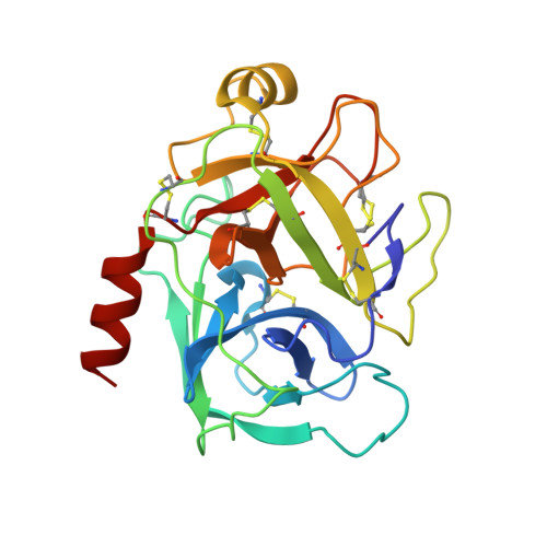
NMR and crystallographic characterization of adventitious borate binding by trypsin.
Transue, T.R., Gabel, S.A., London, R.E.(2006) Bioconjug Chem 17: 300-308
- PubMed: 16536459
- DOI: https://doi.org/10.1021/bc0502210
- Primary Citation of Related Structures:
2A31, 2A32 - PubMed Abstract:
Recent 11B NMR studies of the formation of ternary complexes of trypsin, borate, and S1-binding alcohols revealed evidence for an additional binding interaction external to the enzyme active site. We have explored this binding interaction as a prototypical interaction of borate and boronate ligands with residues on the protein surface. NMR studies of trypsin in which the active site is blocked with leupeptin or with the irreversible inhibitor 4-(2-aminoethyl) benzenesulfonyl fluoride hydrochloride (AEBSF) indicate the existence of a low-affinity borate binding site with an apparent dissociation constant of 97 mM, measured at pH 8.0. Observation of a field-dependent dynamic frequency shift of the (11)B resonance indicates that it corresponds to a complex for which omegatau >> 1. The 0.12 ppm shift difference of the borate resonances measured at 11.75 and 7.05 T, corresponds to a quadrupole coupling constant of 260 kHz. A much larger 2.0 ppm shift is observed in the 11B NMR spectra of trypsin complexed with benzene boronic acid (BBA), leading to a calculated quadrupole coupling constant of 1.1 MHz for this complex. Crystallographic studies identify the second borate binding site as a serine-rich region on the surface of the molecule. Specifically, a complex obtained at pH 10.6 shows a borate ion covalently bonded to the hydroxyl oxygen atoms of Ser164 and Ser167, with additional stabilization coming from two hydrogen-bonding interactions. A similar structure, although with low occupancy (30%), is observed for a trypsin-BBA complex. In this case, the BBA is also observed in the active site, covalently bound in two different conformations to both His57 Nepsilon and Ser195 Ogamma. An analysis of pairwise hydroxyl oxygen distances was able to predict the secondary borate binding site in porcine trypsin, and this approach is potentially useful for prediction of borate binding sites on the surfaces of other proteins. However, the distances between the Ser164/Ser167 Ogamma atoms in all of the reported trypsin crystal structures is significantly greater than the Ogamma distances of 2.2 and 1.9 angstroms observed in the trypsin complexes with borate and BBA, respectively. Thus, the ability of the hydroxyl oxygens to adopt a sufficiently close orientation to allow bidentate ligation is a critical limit on the borate binding affinity of surface-accessible serine/threonine/tyrosine residues.
Organizational Affiliation:
National Institute of Environmental Health Sciences, Laboratory of Structural Biology, Box 12233, Research Triangle Park, North Carolina 27709, USA.


