


Crystal structure of glutamate-1-semialdehyde 2,1-aminomutase from Aeropyrum pernix
Mizutani, H., Kunishima, N.To be published.
Experimental Data Snapshot
Entity ID: 1 | |||||
|---|---|---|---|---|---|
| Molecule | Chains | Sequence Length | Organism | Details | Image |
| Glutamate-1-semialdehyde 2,1-aminomutase | 434 | Aeropyrum pernix | Mutation(s): 0 EC: 5.4.3.8 | 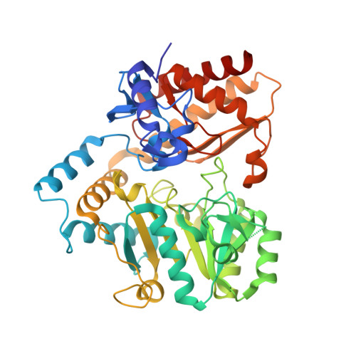 | |
UniProt | |||||
Find proteins for Q9Y9I9 (Aeropyrum pernix (strain ATCC 700893 / DSM 11879 / JCM 9820 / NBRC 100138 / K1)) Explore Q9Y9I9 Go to UniProtKB: Q9Y9I9 | |||||
Entity Groups | |||||
| Sequence Clusters | 30% Identity50% Identity70% Identity90% Identity95% Identity100% Identity | ||||
| UniProt Group | Q9Y9I9 | ||||
Sequence AnnotationsExpand | |||||
| |||||
| Ligands 2 Unique | |||||
|---|---|---|---|---|---|
| ID | Chains | Name / Formula / InChI Key | 2D Diagram | 3D Interactions | |
| PMP Query on PMP | D [auth A], F [auth B], H [auth C] | 4'-DEOXY-4'-AMINOPYRIDOXAL-5'-PHOSPHATE C8 H13 N2 O5 P ZMJGSOSNSPKHNH-UHFFFAOYSA-N |  | ||
| CL Query on CL | E [auth A], G [auth B], I [auth C] | CHLORIDE ION Cl VEXZGXHMUGYJMC-UHFFFAOYSA-M |  | ||
| Length ( Å ) | Angle ( ˚ ) |
|---|---|
| a = 123.39 | α = 90 |
| b = 123.39 | β = 90 |
| c = 356.443 | γ = 120 |
| Software Name | Purpose |
|---|---|
| CNS | refinement |
| HKL-2000 | data collection |
| HKL-2000 | data reduction |
| HKL-2000 | data scaling |
| MOLREP | phasing |
RCSB PDB (citation) is hosted by
RCSB PDB is a member of the

