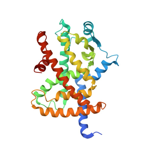
Structural insight into PPARgamma activation through covalent modification with endogenous fatty acids
Waku, T., Shiraki, T., Oyama, T., Fujimoto, Y., Maebara, K., Kamiya, N., Jingami, H., Morikawa, K.(2009) J Mol Biol 385: 188-199
- PubMed: 18977231
- DOI: https://doi.org/10.1016/j.jmb.2008.10.039
- Primary Citation of Related Structures:
2ZK0, 2ZK1, 2ZK2, 2ZK3, 2ZK4, 2ZK5 - PubMed Abstract:
Peroxisome proliferator-activated receptor (PPAR) gamma is a nuclear receptor that regulates lipid homeostasis, and several fatty acid metabolites have been identified as PPARgamma ligands. Here, we present four crystal structures of the PPARgamma ligand binding domain (LBD) covalently bound to endogenous fatty acids via a unique cysteine, which is reportedly critical for receptor activation. The structure analyses of the LBD complexed with 15-deoxy-Delta(12,14)-prostaglandin J(2) (15d-PGJ(2)) revealed that the covalent binding of 15d-PGJ(2) induced conformational changes in the loop region following helix H2', and rearrangements of the side-chain network around the created covalent bond in the LBD. Point mutations of these repositioned residues on the loop and helix H3 almost completely abolished PPARgamma activation by 15d-PGJ(2), indicating that the observed structural alteration may be crucial for PPARgamma activation by the endogenous fatty acid. To address the issue of partial agonism of endogenous PPARgamma ligands, we took advantage of a series of oxidized eicosatetraenoic acids (oxoETEs) as covalently bound ligands to PPARgamma. Despite similar structural and chemical properties, these fatty acids exhibited distinct degrees of transcriptional activity. Crystallographic studies, using two of the oxoETE/PPARgamma LBD complexes, revealed that transcriptional strength of each oxoETE is associated with the difference in the loop conformation, rather than the interaction between each ligand and helix H12. These results suggest that the loop conformation may be responsible for the modulation of PPARgamma activity. Based on these results, we identified novel agonists covalently bound to PPARgamma by in silico screening and a cell-based assay. Our crystallographic study of LBD complexed with nitro-233 demonstrated that the expected covalent bond is indeed formed between this newly identified agonist and the cysteine. This study presents the structural basis for the activation and modulation mechanism of PPARgamma through covalent modification with endogenous fatty acids.
Organizational Affiliation:
Takara Bio Endowed Division, Department of Biomolecular Recognition, Institute for Protein Research, Osaka University, Open Laboratories of Advanced Bioscience and Biotechnology, Suita, Osaka, Japan.