2Z9O

Crystal structure of the dimeric form of RepE in complex with the repE operator DNA


Experimental Data Snapshot

  • Method: X-RAY DIFFRACTION
  • Resolution: 3.14 Å
  • R-Value Free: 0.313 
  • R-Value Work: 0.266 
  • R-Value Observed: 0.266 

wwPDB Validation   3D Report Full Report


This is version 1.2 of the entry. See complete history


Literature

Structural basis for regulation of bifunctional roles in replication initiator protein

Nakamura, A.Wada, C.Miki, K.

(2007) Proc Natl Acad Sci U S A 104: 18484-18489

  • DOI: https://doi.org/10.1073/pnas.0705623104
  • Primary Citation of Related Structures:  
    2Z9O

  • PubMed Abstract: 

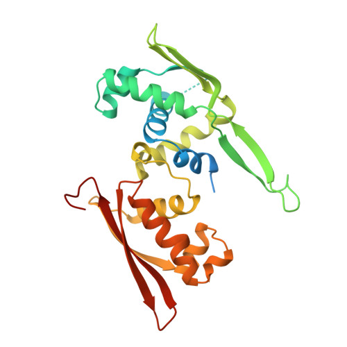
    DNA replication initiator protein RepE stringently regulates F plasmid replication by its two distinct molecular association states. A predominant dimer functions as an autogenous repressor, whereas monomers act as replication initiators, and the dimer requires actions of the DnaK molecular chaperone system for monomerization. The structure of the monomeric form is known, whereas the dimeric structure and structural details of the dimer-to-monomer conversion have been unclear. Here we present the crystal structure of the RepE dimer in complex with the repE operator DNA. The dimerization interface is mainly formed by intermolecular beta-sheets with several key interactions of charged residues. The conformations of the internal N- and C-terminal domains are conserved between the dimer and monomer, whereas the relative domain orientations are strikingly different, allowing for an efficient oligomeric transition of dual-functional RepE. This domain relocation accompanies secondary structural changes in the linker connecting the two domains, and the linker is included in plausible DnaK/DnaJ-binding regions. These findings suggest an activation mechanism for F plasmid replication by RepE monomerization, which is induced and mediated by the DnaK system.


  • Organizational Affiliation

    Department of Chemistry, Graduate School of Science, Kyoto University, Sakyo-ku, Kyoto 606-8502, Japan.


Macromolecules

Find similar proteins by:  (by identity cutoff)  |  3D Structure
Entity ID: 3
MoleculeChains Sequence LengthOrganismDetailsImage
Replication initiation proteinC [auth A],
D [auth B]
266Escherichia coli K-12Mutation(s): 0 
Gene Names: repE
UniProt
Find proteins for P03856 (Escherichia coli (strain K12))
Explore P03856 
Go to UniProtKB:  P03856
Entity Groups  
Sequence Clusters30% Identity50% Identity70% Identity90% Identity95% Identity100% Identity
UniProt GroupP03856
Sequence Annotations
Expand
  • Reference Sequence
Find similar nucleic acids by:  (by identity cutoff)  |  3D Structure
Entity ID: 1
MoleculeChains LengthOrganismImage
DNA (33-MER)A [auth C]33N/A
Sequence Annotations
Expand
  • Reference Sequence
Find similar nucleic acids by:  (by identity cutoff)  |  3D Structure
Entity ID: 2
MoleculeChains LengthOrganismImage
DNA (33-MER)B [auth D]33N/A
Sequence Annotations
Expand
  • Reference Sequence
Experimental Data & Validation

Experimental Data

  • Method: X-RAY DIFFRACTION
  • Resolution: 3.14 Å
  • R-Value Free: 0.313 
  • R-Value Work: 0.266 
  • R-Value Observed: 0.266 
  • Space Group: P 1 21 1
Unit Cell:
Length ( Å )Angle ( ˚ )
a = 60.729α = 90
b = 99.316β = 108.55
c = 95.003γ = 90
Software Package:
Software NamePurpose
CNSrefinement
HKL-2000data reduction
SCALEPACKdata scaling
MOLREPphasing

Structure Validation

View Full Validation Report


Entry History 

Deposition Data

Revision History  (Full details and data files)

  • Version 1.0: 2007-11-27
    Type: Initial release
  • Version 1.1: 2011-07-13
    Changes: Source and taxonomy, Version format compliance
  • Version 1.2: 2023-11-01
    Changes: Data collection, Database references, Refinement description