2YNJ

GroEL at sub-nanometer resolution by Constrained Single Particle Tomography


Experimental Data Snapshot

  • Method: ELECTRON MICROSCOPY
  • Resolution: 8.40 Å
  • Aggregation State: PARTICLE 
  • Reconstruction Method: SINGLE PARTICLE 

wwPDB Validation   3D Report Full Report


This is version 1.2 of the entry. See complete history


Literature

Protein Secondary Structure Determination by Constrained Single-Particle Cryo-Electron Tomography

Bartesaghi, A.Lecumberry, F.Sapiro, G.Subramaniam, S.

(2012) Structure 20: 2003

  • DOI: https://doi.org/10.1016/j.str.2012.10.016
  • Primary Citation of Related Structures:  
    2YNJ

  • PubMed Abstract: 

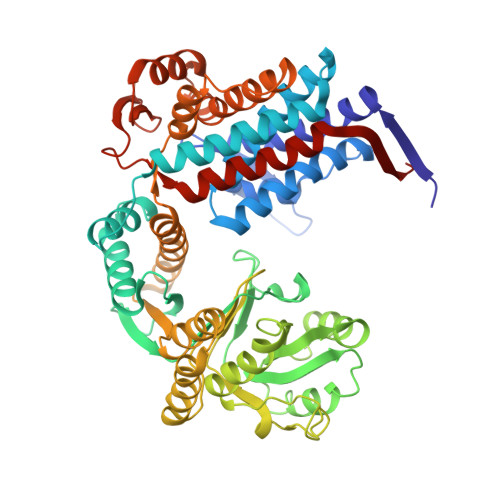
    Cryo-electron microscopy (cryo-EM) is a powerful technique for 3D structure determination of protein complexes by averaging information from individual molecular images. The resolutions that can be achieved with single-particle cryo-EM are frequently limited by inaccuracies in assigning molecular orientations based solely on 2D projection images. Tomographic data collection schemes, however, provide powerful constraints that can be used to more accurately determine molecular orientations necessary for 3D reconstruction. Here, we propose "constrained single-particle tomography" as a general strategy for 3D structure determination in cryo-EM. A key component of our approach is the effective use of images recorded in tilt series to extract high-resolution information and correct for the contrast transfer function. By incorporating geometric constraints into the refinement to improve orientational accuracy of images, we reduce model bias and overrefinement artifacts and demonstrate that protein structures can be determined at resolutions of ∼8 Å starting from low-dose tomographic tilt series.


  • Organizational Affiliation

    Laboratory of Cell Biology, Center for Cancer Research, National Cancer Institute, National Institutes of Health, Bethesda, MD 20892, USA.


Macromolecules
Find similar proteins by:  (by identity cutoff)  |  3D Structure
Entity ID: 1
MoleculeChains Sequence LengthOrganismDetailsImage
60 KDA CHAPERONIN
A, B, C, D, E
A, B, C, D, E, F, G, H, I, J, K, L, M, N
524Escherichia coli UTI89Mutation(s): 0 
UniProt
Find proteins for Q1R3B6 (Escherichia coli (strain UTI89 / UPEC))
Explore Q1R3B6 
Go to UniProtKB:  Q1R3B6
Entity Groups  
Sequence Clusters30% Identity50% Identity70% Identity90% Identity95% Identity100% Identity
UniProt GroupQ1R3B6
Sequence Annotations
Expand
  • Reference Sequence
Small Molecules
Ligands 3 Unique
IDChains Name / Formula / InChI Key2D Diagram3D Interactions
AGS
Query on AGS

Download Ideal Coordinates CCD File 
AA [auth C]
CC [auth L]
EB [auth H]
GA [auth D]
IC [auth M]
AA [auth C],
CC [auth L],
EB [auth H],
GA [auth D],
IC [auth M],
KB [auth I],
MA [auth E],
O [auth A],
OC [auth N],
QB [auth J],
SA [auth F],
U [auth B],
WB [auth K],
YA [auth G]
PHOSPHOTHIOPHOSPHORIC ACID-ADENYLATE ESTER
C10 H16 N5 O12 P3 S
NLTUCYMLOPLUHL-KQYNXXCUSA-N
TL
Query on TL

Download Ideal Coordinates CCD File 
AB [auth G]
BA [auth C]
BB [auth G]
BC [auth K]
CA [auth C]
AB [auth G],
BA [auth C],
BB [auth G],
BC [auth K],
CA [auth C],
DA [auth C],
DB [auth G],
DC [auth L],
EC [auth L],
FA [auth C],
FB [auth H],
FC [auth L],
GB [auth H],
HA [auth D],
HB [auth H],
HC [auth L],
IA [auth D],
JA [auth D],
JB [auth H],
JC [auth M],
KC [auth M],
LA [auth D],
LB [auth I],
LC [auth M],
MB [auth I],
NA [auth E],
NB [auth I],
NC [auth M],
OA [auth E],
P [auth A],
PA [auth E],
PB [auth I],
PC [auth N],
Q [auth A],
QC [auth N],
R [auth A],
RA [auth E],
RB [auth J],
RC [auth N],
SB [auth J],
T [auth A],
TA [auth F],
TB [auth J],
TC [auth N],
UA [auth F],
V [auth B],
VA [auth F],
VB [auth J],
W [auth B],
X [auth B],
XA [auth F],
XB [auth K],
YB [auth K],
Z [auth B],
ZA [auth G],
ZB [auth K]
THALLIUM (I) ION
Tl
ZLUSCZLCHQSJRU-UHFFFAOYSA-N
MG
Query on MG

Download Ideal Coordinates CCD File 
AC [auth K]
CB [auth G]
EA [auth C]
GC [auth L]
IB [auth H]
AC [auth K],
CB [auth G],
EA [auth C],
GC [auth L],
IB [auth H],
KA [auth D],
MC [auth M],
OB [auth I],
QA [auth E],
S [auth A],
SC [auth N],
UB [auth J],
WA [auth F],
Y [auth B]
MAGNESIUM ION
Mg
JLVVSXFLKOJNIY-UHFFFAOYSA-N
Experimental Data & Validation

Experimental Data

  • Method: ELECTRON MICROSCOPY
  • Resolution: 8.40 Å
  • Aggregation State: PARTICLE 
  • Reconstruction Method: SINGLE PARTICLE 
EM Software:
TaskSoftware PackageVersion
RECONSTRUCTIONFREALIGN

Structure Validation

View Full Validation Report


Entry History 

Deposition Data

Revision History  (Full details and data files)

  • Version 1.0: 2012-12-12
    Type: Initial release
  • Version 1.1: 2012-12-19
    Changes: Database references
  • Version 1.2: 2017-08-23
    Changes: Data collection, Refinement description