2Y7B

Crystal structure of the PH domain of human Actin-binding protein anillin ANLN


Experimental Data Snapshot

  • Method: X-RAY DIFFRACTION
  • Resolution: 1.90 Å
  • R-Value Free: 0.262 
  • R-Value Work: 0.192 
  • R-Value Observed: 0.195 

wwPDB Validation   3D Report Full Report


This is version 1.3 of the entry. See complete history


Literature

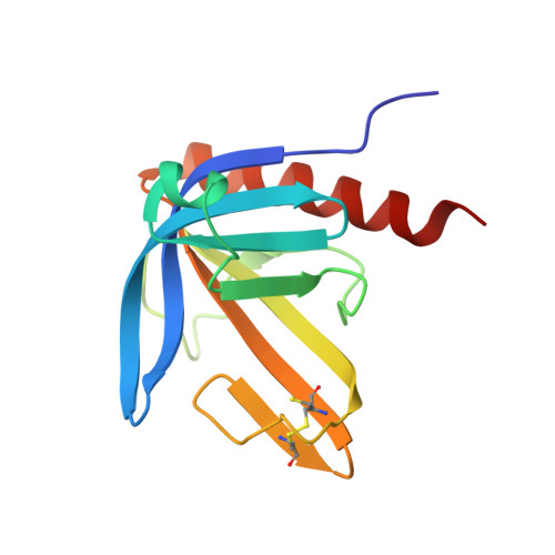
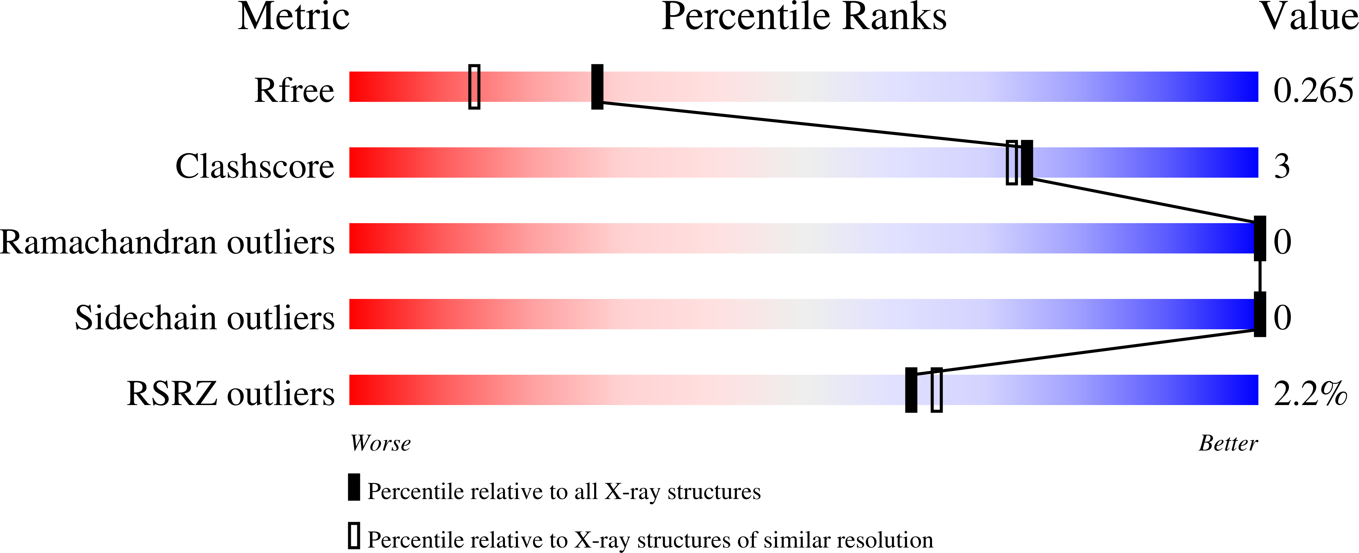
Crystal Structure of the Ph Domain of Human Actin-Binding Protein Anillin Anln

Vollmar, M.Wang, J.Krojer, T.Elkins, J.Filippakopoulos, P.Ugochukwu, E.Cocking, R.von Delft, F.Bountra, C.Arrowsmith, C.H.Weigelt, J.Edwards, A.Knapp, S.

To be published.

Macromolecules
Find similar proteins by:  (by identity cutoff)  |  3D Structure
Entity ID: 1
MoleculeChains Sequence LengthOrganismDetailsImage
ACTIN-BINDING PROTEIN ANILLIN134Homo sapiensMutation(s): 0 
UniProt & NIH Common Fund Data Resources
Find proteins for Q9NQW6 (Homo sapiens)
Explore Q9NQW6 
Go to UniProtKB:  Q9NQW6
PHAROS:  Q9NQW6
GTEx:  ENSG00000011426 
Entity Groups  
Sequence Clusters30% Identity50% Identity70% Identity90% Identity95% Identity100% Identity
UniProt GroupQ9NQW6
Sequence Annotations
Expand
  • Reference Sequence
Experimental Data & Validation

Experimental Data

  • Method: X-RAY DIFFRACTION
  • Resolution: 1.90 Å
  • R-Value Free: 0.262 
  • R-Value Work: 0.192 
  • R-Value Observed: 0.195 
  • Space Group: P 21 21 21
Unit Cell:
Length ( Å )Angle ( ˚ )
a = 34.3α = 90
b = 59.417β = 90
c = 64.717γ = 90
Software Package:
Software NamePurpose
REFMACrefinement
XDSdata reduction
SCALAdata scaling
PHASERphasing

Structure Validation

View Full Validation Report


Entry History 

Revision History  (Full details and data files)

  • Version 1.0: 2011-03-30
    Type: Initial release
  • Version 1.1: 2012-12-05
    Changes: Database references, Structure summary, Version format compliance
  • Version 1.2: 2018-01-24
    Changes: Database references
  • Version 1.3: 2023-12-20
    Changes: Data collection, Database references, Derived calculations, Other, Refinement description