2Y6X

Structure of Psb27 from Thermosynechococcus elongatus


Experimental Data Snapshot

  • Method: X-RAY DIFFRACTION
  • Resolution: 1.60 Å
  • R-Value Free: 0.238 
  • R-Value Work: 0.190 
  • R-Value Observed: 0.192 

wwPDB Validation   3D Report Full Report


This is version 1.3 of the entry. See complete history


Literature

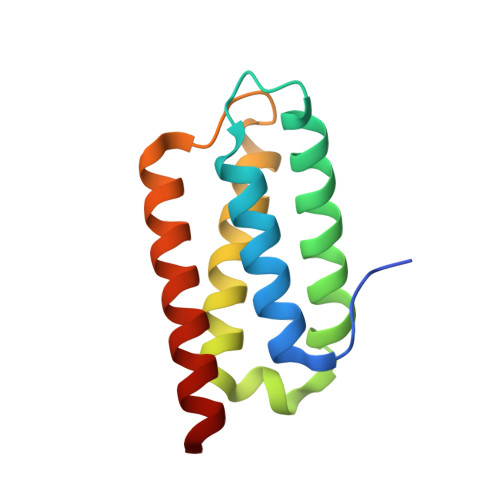
Crystal Structure of the Psb27 Assembly Factor at 1.6A: Implications for Binding to Photosystem II.

Michoux, F.Takasaka, K.Boehm, M.Komenda, J.Nixon, P.J.Murray, J.W.

(2012) Photosynth Res 110: 169

  • DOI: https://doi.org/10.1007/s11120-011-9712-7
  • Primary Citation of Related Structures:  
    2Y6X

  • PubMed Abstract: 

    The biogenesis and oxygen-evolving activity of cyanobacterial Photosystem II (PSII) is dependent on a number of accessory proteins not found in the crystallised dimeric complex. These include Psb27, a small lipoprotein attached to the lumenal side of PSII, which has been assigned a role in regulating the assembly of the Mn(4)Ca cluster catalysing water oxidation. To gain a better understanding of Psb27, we have determined in this study the crystal structure of the soluble domain of Psb27 from Thermosynechococcus elongatus to a resolution of 1.6 Å. The structure is a four-helix bundle, similar to the recently published solution structures of Psb27 from Synechocystis PCC 6803 obtained by nuclear magnetic resonance (NMR) spectroscopy. Importantly, the crystal structure presented here helps us resolve the differences between the NMR-derived structural models. Potential binding sites for Psb27 within PSII are discussed in light of recent biochemical data in the literature.


  • Organizational Affiliation

    Division of Molecular Biosciences, Imperial College London, Wolfson Biochemistry Building, South Kensington Campus, London, SW7 2AZ, UK. franck.michoux@imperial.ac.uk


Macromolecules
Find similar proteins by:  (by identity cutoff)  |  3D Structure
Entity ID: 1
MoleculeChains Sequence LengthOrganismDetailsImage
PHOTOSYSTEM II 11 KD PROTEIN113Thermosynechococcus vestitusMutation(s): 0 
UniProt
Find proteins for Q8DG60 (Thermosynechococcus vestitus (strain NIES-2133 / IAM M-273 / BP-1))
Explore Q8DG60 
Go to UniProtKB:  Q8DG60
Entity Groups  
Sequence Clusters30% Identity50% Identity70% Identity90% Identity95% Identity100% Identity
UniProt GroupQ8DG60
Sequence Annotations
Expand
  • Reference Sequence
Small Molecules
Ligands 1 Unique
IDChains Name / Formula / InChI Key2D Diagram3D Interactions
CL
Query on CL

Download Ideal Coordinates CCD File 
B [auth A]CHLORIDE ION
Cl
VEXZGXHMUGYJMC-UHFFFAOYSA-M
Experimental Data & Validation

Experimental Data

  • Method: X-RAY DIFFRACTION
  • Resolution: 1.60 Å
  • R-Value Free: 0.238 
  • R-Value Work: 0.190 
  • R-Value Observed: 0.192 
  • Space Group: P 21 21 21
Unit Cell:
Length ( Å )Angle ( ˚ )
a = 36.93α = 90
b = 50.82β = 90
c = 53.07γ = 90
Software Package:
Software NamePurpose
REFMACrefinement
MOSFLMdata reduction
SCALAdata scaling
PHASERphasing

Structure Validation

View Full Validation Report


Entry History 

Deposition Data

Revision History  (Full details and data files)

  • Version 1.0: 2012-01-11
    Type: Initial release
  • Version 1.1: 2012-02-15
    Changes: Other
  • Version 1.2: 2019-05-08
    Changes: Data collection, Experimental preparation, Other
  • Version 1.3: 2023-12-20
    Changes: Data collection, Database references, Derived calculations, Other, Refinement description