2Y60

Isopenicillin N synthase with AC-D-methionine


Experimental Data Snapshot

  • Method: X-RAY DIFFRACTION
  • Resolution: 1.40 Å
  • R-Value Free: 0.199 
  • R-Value Work: 0.145 
  • R-Value Observed: 0.147 

wwPDB Validation   3D Report Full Report


Ligand Structure Quality Assessment 


This is version 1.3 of the entry. See complete history


Literature

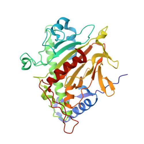
The Crystal Structure of Isopenicillin N Synthase with Delta((L)-Alpha-Aminoadipoyl)-(L)-Cysteinyl-(D)-Methionine Reveals Thioether Coordination to Iron.

Clifton, I.J.Ge, W.Adlington, R.M.Baldwin, J.E.Rutledge, P.J.

(2011) Arch Biochem Biophys 516: 103

  • DOI: https://doi.org/10.1016/j.abb.2011.09.014
  • Primary Citation of Related Structures:  
    2Y60

  • PubMed Abstract: 

    Isopenicillin N synthase (IPNS) catalyses cyclization of δ-(l-α-aminoadipoyl)-l-cysteinyl-d-valine (ACV) to isopenicillin N (IPN), the central step in penicillin biosynthesis. Previous studies have shown that IPNS turns over a wide range of substrate analogues in which the valine residue of its natural substrate is replaced with other amino acids. IPNS accepts and oxidizes numerous substrates that bear hydrocarbon sidechains in this position, however the enzyme is less tolerant of analogues presenting polar functionality in place of the valinyl isopropyl group. We report a new ACV analogue δ-(l-α-aminoadipoyl)-l-cysteinyl-d-methionine (ACM), which incorporates a thioether in place of the valinyl sidechain. ACM has been synthesized using solution phase methods and crystallized with IPNS. A crystal structure has been elucidated for the IPNS:Fe(II):ACM complex at 1.40Å resolution. This structure reveals that ACM binds in the IPNS active site such that the sulfur atom of the methionine thioether binds to iron in the oxygen binding site at a distance of 2.57Å. The sulfur of the cysteinyl thiolate sits 2.36Å from the metal.


  • Organizational Affiliation

    Chemistry Research Laboratory, University of Oxford, UK.


Macromolecules
Find similar proteins by:  (by identity cutoff)  |  3D Structure
Entity ID: 1
MoleculeChains Sequence LengthOrganismDetailsImage
ISOPENICILLIN N SYNTHASE331Aspergillus nidulansMutation(s): 0 
EC: 1.21.3.1
UniProt
Find proteins for P05326 (Emericella nidulans (strain FGSC A4 / ATCC 38163 / CBS 112.46 / NRRL 194 / M139))
Explore P05326 
Go to UniProtKB:  P05326
Entity Groups  
Sequence Clusters30% Identity50% Identity70% Identity90% Identity95% Identity100% Identity
UniProt GroupP05326
Sequence Annotations
Expand
  • Reference Sequence
Experimental Data & Validation

Experimental Data

  • Method: X-RAY DIFFRACTION
  • Resolution: 1.40 Å
  • R-Value Free: 0.199 
  • R-Value Work: 0.145 
  • R-Value Observed: 0.147 
  • Space Group: P 21 21 21
Unit Cell:
Length ( Å )Angle ( ˚ )
a = 46.523α = 90
b = 70.882β = 90
c = 100.923γ = 90
Software Package:
Software NamePurpose
REFMACrefinement
DENZOdata reduction
SCALAdata scaling
REFMACphasing

Structure Validation

View Full Validation Report


Ligand Structure Quality Assessment 


Entry History 

Deposition Data

Revision History  (Full details and data files)

  • Version 1.0: 2012-02-08
    Type: Initial release
  • Version 1.1: 2012-08-15
    Changes: Database references, Non-polymer description
  • Version 1.2: 2019-07-17
    Changes: Data collection
  • Version 1.3: 2023-12-20
    Changes: Data collection, Database references, Derived calculations, Other, Refinement description